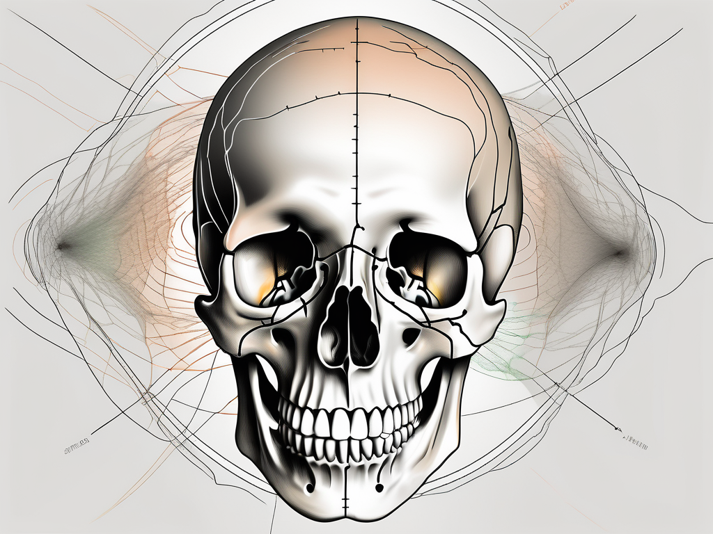
The abducens nerve, also known as cranial nerve VI, is a crucial component of the human nervous system. It plays a vital role in the movement of the eyes, specifically controlling the lateral rectus muscle. This muscle allows the eyes to move laterally, helping us gaze in different directions. To fully comprehend the significance of the abducens nerve, it is essential to understand its anatomy and function.
Understanding the Abducens Nerve
Anatomy of the Abducens Nerve
Located within the cavernous sinus, the abducens nerve originates from the pons and extends towards the eye socket. Emerging from the brainstem, it travels through the superior orbital fissure, a narrow opening in the skull. Once it reaches the eye socket, it innervates the lateral rectus muscle on each side, enabling precise eye movements.
The abducens nerve, also known as the sixth cranial nerve, is a vital component of the intricate network of nerves that control eye movement. It is one of the twelve cranial nerves that emerge directly from the brain, making it an essential link between the central nervous system and the eyes.
As the abducens nerve makes its way from the pons to the eye socket, it navigates through a complex pathway, ensuring the transmission of signals necessary for proper eye function. This intricate journey highlights the remarkable precision of the human body’s anatomical design.
Function of the Abducens Nerve
The primary function of the abducens nerve is to control the lateral movement of the eyes. Its coordinated action with other cranial nerves ensures that our eyes work harmoniously, enabling us to shift our gaze from one point to another.
When the abducens nerve is functioning correctly, both eyes can move outward simultaneously, facilitating binocular vision. This coordinated movement is essential for activities such as reading, driving, and even everyday tasks like crossing the road.
Moreover, the abducens nerve plays a crucial role in maintaining eye alignment. It helps prevent conditions such as strabismus, commonly known as crossed eyes, by ensuring that both eyes move in unison. This precise control allows for optimal visual perception and depth perception, enhancing our overall visual experience.
Furthermore, the abducens nerve’s function extends beyond simple eye movements. It also contributes to the complex process of eye tracking, which involves following objects with our eyes as they move. This ability is essential for various tasks, including reading, playing sports, and interacting with our environment.
In summary, the abducens nerve’s anatomy and function are intricately linked, showcasing the remarkable complexity and precision of the human body. Its role in controlling eye movements and maintaining eye alignment highlights its significance in our everyday lives. Understanding the abducens nerve allows us to appreciate the incredible mechanisms that enable us to see and navigate the world around us.
The Concept of Foramen in Human Anatomy
Definition and Role of Foramen
In the vast landscape of human anatomy, a foramen is a vital structure. Derived from Latin, the word “foramen” means an opening or hole. In the context of the body, a foramen refers to a natural passageway that allows nerves, blood vessels, and connective tissues to pass through or connect different anatomical regions.
Foramen not only provide passage but also serve as protective structures for various anatomical components. They play a critical role in ensuring the optimal functioning and communication between different parts of the body.
Foramen can be found throughout the body, from the skull to the pelvis, and even in the extremities. These openings are strategically positioned to facilitate the flow of essential structures, such as nerves and blood vessels, to their intended destinations.
One example of a foramen is the foramen lacerum, located in the base of the skull. This triangular-shaped opening allows for the passage of important structures, including the internal carotid artery and the greater petrosal nerve. Without the foramen lacerum, these vital structures would not be able to reach their respective destinations, leading to potential complications and impairments.
Different Types of Foramen
The human body has numerous foramen, each serving a specific purpose in facilitating bodily functions. Some notable examples include the foramen magnum, which allows the spinal cord to connect with the brain, and the foramen ovale, which provides a pathway for vessels and nerves in the heart.
When it comes to the abducens nerve, there is a unique foramen that it traverses on its journey to the eye socket. This foramen, known as the superior orbital fissure, is a narrow opening located in the sphenoid bone. It serves as a crucial pathway for the abducens nerve, allowing it to reach the muscles responsible for the lateral movement of the eye. Without the superior orbital fissure, the abducens nerve would be unable to fulfill its role, resulting in impaired eye movement and coordination.
Another fascinating example of a foramen is the foramen rotundum, found in the sphenoid bone as well. This circular opening allows for the passage of the maxillary nerve, one of the three branches of the trigeminal nerve. The maxillary nerve is responsible for providing sensory innervation to the upper teeth, palate, and nasal cavity. Through the foramen rotundum, the maxillary nerve can reach its target areas, ensuring proper sensation and function.
It is important to note that foramen can vary in size and shape, depending on their location and the structures they accommodate. Some foramen may be small and narrow, while others can be larger and more spacious. The diversity in foramen characteristics reflects the complexity and intricacy of human anatomy.
The Journey of the Abducens Nerve
Origin and Pathway of the Abducens Nerve
As mentioned earlier, the abducens nerve originates from the pons, which is a crucial part of the brainstem. The pons, located in the upper part of the brainstem, serves as a bridge connecting various regions of the brain. It plays a vital role in relaying signals between the cerebral cortex and the spinal cord.
From its origin in the pons, the abducens nerve embarks on a fascinating journey, traversing a specific path through the intricate network of structures within the skull. This journey is essential for the nerve to reach its ultimate destination and fulfill its crucial role in eye movement.
The abducens nerve passes through a small opening in the skull called the superior orbital fissure. This foramen, located in the sphenoid bone, connects the middle cranial fossa to the orbit, offering a direct pathway for the abducens nerve to reach the lateral rectus muscle in the eye socket.
As the abducens nerve makes its way through the superior orbital fissure, it encounters a complex arrangement of structures. Surrounding the nerve are various blood vessels, connective tissues, and other cranial nerves, creating a densely packed environment within the confines of the skull.
The Specific Foramen for the Abducens Nerve
Of the many foramina present in the human body, the superior orbital fissure is the designated passageway for the abducens nerve. This narrow opening, located at the junction of the greater and lesser wings of the sphenoid bone, allows the abducens nerve to exit the skull and innervate the lateral rectus muscle, enabling lateral eye movements.
The precise anatomical arrangement and pathway of the abducens nerve through the superior orbital fissure contribute to its critical role in eye movement. The nerve’s passage through this specific foramen ensures its protection and provides a direct route to its target muscle.
It is fascinating to consider the intricate nature of the human body, where even the smallest structures like foramina play a significant role in facilitating the functions of vital nerves such as the abducens nerve. The journey of this nerve through the superior orbital fissure showcases the remarkable precision and complexity of our anatomy.
Implications of Abducens Nerve Passage
The abducens nerve plays a crucial role in controlling eye movement. It is responsible for the lateral movement of the eyes, allowing us to shift our gaze from side to side. Any disruption or damage to the abducens nerve can have significant clinical implications, affecting our ability to control eye movements effectively.
Conditions that affect the passage of the abducens nerve through the superior orbital fissure, a narrow opening in the skull, can result in various symptoms and impairments. These symptoms can vary depending on the extent and location of the damage.
Clinical Significance of Abducens Nerve Passage
One of the most common clinical signs associated with abducens nerve dysfunction is double vision, also known as diplopia. When the abducens nerve is affected, the eyes may not be able to coordinate properly, leading to overlapping images. This can significantly impact daily activities such as reading, driving, and even simple tasks like walking.
In addition to double vision, individuals with abducens nerve dysfunction may experience a decreased ability to move their eyes laterally. This limitation in eye movement can make it challenging to focus on objects located to the side, affecting peripheral vision and overall visual perception.
Misalignment of the eyes, known as strabismus, is another common clinical sign of abducens nerve dysfunction. In this condition, one eye may deviate inward or outward, causing an imbalance in the alignment of the eyes. Strabismus can lead to difficulties with depth perception and may cause social and psychological challenges due to the noticeable difference in eye appearance.
While these symptoms can occur for various reasons, it is crucial to consult with a healthcare professional for a precise diagnosis and appropriate treatment options. The underlying cause of abducens nerve dysfunction can vary, and a thorough evaluation is necessary to determine the best course of action.
Disorders Related to Abducens Nerve Passage
Several disorders can impact the abducens nerve’s passage through the superior orbital fissure, thereby affecting eye movement. Tumors, both benign and malignant, can exert pressure on the nerve, causing compression and impairing its function. Traumatic head injuries, such as skull fractures or severe blows to the head, can also damage the abducens nerve, leading to dysfunction.
Infections, such as sinusitis or meningitis, can also affect the abducens nerve’s passage through the superior orbital fissure. Inflammatory processes associated with these infections can cause swelling and compression of the nerve, resulting in symptoms of abducens nerve dysfunction.
Furthermore, genetic abnormalities can contribute to abducens nerve dysfunction. Certain inherited conditions, such as Duane syndrome or congenital cranial dysinnervation disorders, can affect the development and function of the abducens nerve, leading to various eye movement abnormalities.
When faced with any changes in eye movement or vision, it is always advisable to seek medical attention promptly. An accurate diagnosis can help determine the underlying cause of the symptoms and guide appropriate treatment strategies. Early intervention is crucial in managing abducens nerve dysfunction and minimizing its impact on daily life.
Frequently Asked Questions about Abducens Nerve and Foramen
Common Misconceptions about Abducens Nerve and Foramen
There are various misconceptions surrounding the abducens nerve and its relation to foramen. One common misconception is that any disruption to the abducens nerve’s passage through the superior orbital fissure automatically results in vision loss. While it is true that abducens nerve dysfunction can cause visual disturbances, complete vision loss is not a common manifestation.
However, it is important to note that the abducens nerve plays a vital role in eye movement and coordination. This cranial nerve innervates the lateral rectus muscle, which is responsible for the outward movement of the eye. Any disruption to the abducens nerve’s function can lead to a condition called abducens nerve palsy, characterized by an inability to move the affected eye laterally.
Another misconception is that all foramen have identical structures and functions. In reality, each foramen has specific anatomical characteristics and serves distinct purposes in different areas of the body.
For example, the superior orbital fissure, through which the abducens nerve passes, is a bony opening located in the skull. It is situated between the greater and lesser wings of the sphenoid bone and serves as a passageway for several important structures, including the abducens nerve, oculomotor nerve, trochlear nerve, and ophthalmic branch of the trigeminal nerve.
On the other hand, the foramen magnum, located at the base of the skull, is the largest foramen in the human body. It allows for the passage of the spinal cord, connecting the brain to the rest of the body. Additionally, the foramen ovale, found in the sphenoid bone, permits the passage of the mandibular branch of the trigeminal nerve, which is responsible for sensory innervation of the face.
Expert Answers to Your Questions
If you have any questions or concerns about the abducens nerve, its passage through the superior orbital fissure, or any other related topic, it is best to consult with a healthcare professional. Qualified medical experts can provide accurate information, address individual concerns, and offer appropriate guidance based on your specific circumstances.
Remember, this article provides comprehensive information but should not be taken as medical advice. It is always essential to seek professional help for reliable diagnosis and treatment of any health-related issues you may be experiencing.
In conclusion, the abducens nerve passes through the superior orbital fissure on its journey from the pons to the eye socket. This anatomical pathway is crucial for proper eye movement and coordination. Understanding the complex interplay between the abducens nerve and foramen is vital in comprehending its significance and potential clinical implications.
Furthermore, the abducens nerve is part of the cranial nerve system, specifically the sixth cranial nerve. It originates from the pons, a region of the brainstem, and travels through the superior orbital fissure to reach the eye socket. Along its course, the abducens nerve interacts with various structures, including blood vessels and other cranial nerves.
Disorders affecting the abducens nerve can have different causes, ranging from trauma and infections to tumors and vascular abnormalities. These conditions can lead to abducens nerve palsy, resulting in the inability to move the affected eye laterally. Prompt diagnosis and appropriate management are crucial in such cases to prevent further complications and optimize visual function.
If you have any concerns or questions about the abducens nerve or foramen, do not hesitate to consult with a healthcare professional to ensure accurate diagnosis and appropriate management.

Leave a Reply