The abducens nerve, also known as the sixth cranial nerve, plays a crucial role in eye movement. It is responsible for controlling the lateral rectus muscle, which allows the eye to move outward. Assessing the abducens nerve is essential in diagnosing and managing various conditions that can affect its function. In this article, we will delve into the understanding of the abducens nerve, discuss the signs and symptoms of abducens nerve damage, explore diagnostic procedures for assessment, interpret assessment results, and explore treatment options. It is important to note that this article aims to provide information and should not be considered as medical advice. If you suspect any issues with your abducens nerve, we strongly recommend consulting with a medical professional for a proper evaluation and treatment plan.
Understanding the Abducens Nerve
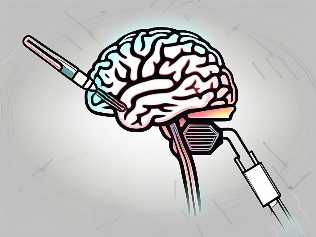
The abducens nerve, also known as the sixth cranial nerve, is a crucial component of the intricate network that controls eye movement. It originates from the pons, a region in the brainstem responsible for relaying signals between the brain and the rest of the body. Specifically, the abducens nerve consists of motor fibers that innervate the lateral rectus muscle, one of the six extraocular muscles that control eye movement.
Before delving into the significance of assessing the abducens nerve, it is essential to comprehend its anatomy and function. By gaining a comprehensive understanding of this nerve, healthcare professionals can better diagnose and treat conditions that may affect its normal functioning.
Anatomy of the Abducens Nerve
The abducens nerve emerges from the brainstem and takes a remarkable journey through the cavernous sinus, a space located behind the eye socket. This intricate pathway exposes the nerve to potential vulnerabilities, making it susceptible to injury or compression. The cavernous sinus, a complex structure filled with blood vessels and nerves, acts as a protective conduit for the abducens nerve as it traverses towards its destination.
Upon exiting the cavernous sinus, the abducens nerve continues its course and reaches the lateral rectus muscle. This muscle, located on the outer side of the eye, plays a vital role in horizontal eye movement. When the abducens nerve stimulates the lateral rectus muscle, it contracts, allowing the eye to move away from the midline. This coordinated movement of both eyes ensures binocular vision, enabling us to perceive the world around us with depth and accuracy.
Function of the Abducens Nerve
The primary function of the abducens nerve is to control the lateral rectus muscle, facilitating the outward movement of the eye. This movement is essential for various visual tasks, such as tracking moving objects or scanning the environment. When the abducens nerve is functioning optimally, both eyes work together harmoniously, allowing us to maintain binocular vision and perceive the world in three dimensions.
However, any disruption in the abducens nerve’s normal functioning can lead to a range of symptoms and visual impairments. Conditions such as abducens nerve palsy, which occurs when the nerve is damaged or compressed, can result in a weakened or paralyzed lateral rectus muscle. This can cause a significant limitation in eye movement, leading to double vision, difficulty focusing, and an overall decrease in visual acuity.
Assessing the abducens nerve’s integrity is crucial in diagnosing and managing various neurological and ophthalmological conditions. Healthcare professionals employ a variety of techniques, including eye movement examinations, imaging studies, and electrophysiological tests, to evaluate the function of this vital nerve.
In conclusion, the abducens nerve plays a pivotal role in controlling eye movement and maintaining optimal visual perception. Its anatomy and function are intricately intertwined, and any disruption in its normal functioning can have profound effects on our ability to see and interpret the world around us. By understanding the complexities of the abducens nerve, healthcare professionals can provide accurate diagnoses and develop effective treatment strategies for patients with conditions affecting this vital cranial nerve.
The Importance of Assessing the Abducens Nerve
Evaluating the abducens nerve is crucial in identifying potential issues with eye movement and ensuring overall neurological health.
The abducens nerve, also known as the sixth cranial nerve, is responsible for the innervation of the lateral rectus muscle, which controls the movement of the eye away from the midline. This nerve plays a key role in facilitating horizontal eye movement, allowing us to track moving objects and scan the environment. Without the proper functioning of the abducens nerve, individuals may experience difficulties in smoothly coordinating their eye movements, leading to impaired visual tracking and reduced visual acuity.
Proper assessment of the abducens nerve can provide valuable insights into an individual’s ability to perform smooth and accurate eye movements, which is essential for various daily activities. For example, when reading a book or following a moving object, the eyes need to move in a coordinated manner to maintain focus and gather visual information effectively. Any disruption in the functioning of the abducens nerve can lead to eye misalignment, resulting in double vision or a reduced field of vision.
Implications for Neurological Health
Assessing the abducens nerve can also help identify underlying neurological conditions. Damage or dysfunction of the nerve can result from various causes, including trauma, tumors, infections, and neurological disorders. For instance, a head injury can cause direct damage to the abducens nerve, leading to impaired eye movement and diplopia (double vision). In some cases, tumors or infections in the brain can exert pressure on the nerve, compromising its function.
By identifying and addressing these issues early on, medical professionals can potentially prevent further complications and ensure the most appropriate treatment approach. For example, if a patient presents with sudden-onset double vision, a thorough assessment of the abducens nerve can help determine the underlying cause. Prompt diagnosis and intervention can prevent permanent damage and improve the patient’s quality of life.
In addition to assessing the abducens nerve, a comprehensive neurological evaluation may involve testing other cranial nerves, such as the oculomotor and trochlear nerves, to assess the overall integrity of the visual system. This evaluation may include assessing pupillary reflexes, eye movements in different directions, and the coordination of eye movements with head movements. By examining the function of multiple cranial nerves, healthcare professionals can gain a more comprehensive understanding of the patient’s neurological health and make informed decisions regarding their care.
Signs and Symptoms of Abducens Nerve Damage
Abducens nerve damage can manifest in both physical and vision-related symptoms. Understanding these symptoms can help individuals identify and seek appropriate medical attention.
Physical Symptoms
Physically, individuals with abducens nerve damage may experience difficulties moving their eyes laterally. This can lead to a condition called “strabismus,” where the affected eye deviates inward, making it challenging to align both eyes simultaneously. Strabismus not only affects the appearance of the eyes but can also impact depth perception and coordination.
In addition to strabismus, some individuals may also experience eye pain, headaches, or discomfort. These symptoms can be particularly bothersome when trying to focus their gaze in a specific direction. The pain and discomfort may vary in intensity, ranging from mild to severe, depending on the extent of the nerve damage.
Furthermore, abducens nerve damage can also cause weakness in the muscles responsible for eye movement. This weakness may result in difficulties in tracking moving objects or following a moving target. Individuals may find it challenging to perform tasks that require precise eye movements, such as reading, playing sports, or even watching television.
Vision-Related Symptoms
When the abducens nerve is affected, individuals may experience double vision, also known as “diplopia.” This occurs because the eyes are not properly aligned, causing the brain to receive conflicting visual information from each eye. Diplopia can be disruptive and interfere with daily activities such as reading, driving, and even walking.
The severity of diplopia can vary depending on the extent of the nerve damage. Some individuals may only experience double vision when looking in a specific direction, while others may have constant double vision regardless of eye movement. This can significantly impact an individual’s quality of life, making simple tasks more challenging and frustrating.
In addition to double vision, abducens nerve damage can also affect visual acuity. Individuals may notice a decrease in their ability to see objects clearly, especially when looking at things far away. This blurred vision can make it difficult to read signs, recognize faces, or even watch television from a comfortable distance.
Moreover, individuals with abducens nerve damage may also experience eye strain and fatigue. The eyes may feel tired and achy, especially after prolonged periods of visual concentration. This can make it challenging to engage in activities that require sustained visual attention, such as reading, working on a computer, or driving for long distances.
It is important to note that the signs and symptoms of abducens nerve damage may vary from person to person. Some individuals may only experience mild symptoms, while others may have more severe impairments. Seeking medical evaluation and treatment can help manage these symptoms and improve overall visual function.
Diagnostic Procedures for Abducens Nerve Assessment
Several diagnostic procedures can be employed to assess the abducens nerve. It is important to accurately diagnose any dysfunction or abnormalities in order to provide appropriate treatment and management.
Clinical Examination Techniques
During a clinical examination, the medical professional may employ various techniques to evaluate the abducens nerve. These techniques involve assessing the function and integrity of the nerve, as well as determining the extent of any dysfunction.
One of the primary aspects of a clinical examination is the evaluation of eye movement. The medical professional will carefully observe the patient’s eye movements, looking for any abnormalities or limitations. They may ask the patient to follow a moving object with their eyes or perform specific eye movements to assess the functionality of the abducens nerve.
In addition to evaluating eye movement, the medical professional will also assess visual acuity. This involves testing the patient’s ability to see clearly and distinguish objects at various distances. By evaluating visual acuity, the medical professional can gain further insight into the function of the abducens nerve.
Furthermore, additional tests may be performed during the clinical examination to provide a comprehensive assessment of the abducens nerve. These tests may include assessing eye alignment, measuring the range of eye movement, and evaluating the presence of diplopia (double vision). These tests help to identify any specific abnormalities or dysfunctions related to the abducens nerve.
Imaging Techniques
In some cases, medical imaging techniques such as magnetic resonance imaging (MRI) or computed tomography (CT) scans may be utilized to visualize the abducens nerve and surrounding structures. These imaging techniques provide detailed images of the brain, cranial nerves, and other relevant anatomical structures.
By using MRI or CT scans, medical professionals can identify potential structural abnormalities or lesions affecting the abducens nerve. These abnormalities may include tumors, inflammation, or other conditions that can impede the normal function of the nerve.
During the imaging procedure, the patient will be positioned inside a machine that uses strong magnetic fields or X-rays to create detailed images. These images are then analyzed by radiologists or other specialists to identify any abnormalities or irregularities that may be affecting the abducens nerve.
Overall, the combination of clinical examination techniques and imaging procedures allows medical professionals to thoroughly assess the abducens nerve and determine the underlying cause of any dysfunction or abnormalities. This comprehensive evaluation is crucial in developing an appropriate treatment plan and ensuring the best possible outcomes for patients.
Interpreting Assessment Results
Interpreting the abducens nerve assessment results is essential in determining the presence and severity of any abnormalities.
The abducens nerve assessment is a crucial diagnostic tool used by healthcare professionals to evaluate the functionality of the abducens nerve, also known as the sixth cranial nerve. This nerve plays a vital role in eye movement, specifically in the lateral movement of the eye.
During the assessment, the healthcare professional carefully observes the patient’s eye movements, looking for any signs of abnormality. They assess the patient’s ability to move their eyes laterally, ensuring that both eyes move in synchrony and without any restrictions.
Normal vs. Abnormal Findings
A normal abducens nerve assessment typically demonstrates intact eye movement, normal eye alignment, and absence of diplopia. When the abducens nerve is functioning properly, the patient’s eyes move smoothly and symmetrically, allowing them to focus on objects in their peripheral vision effortlessly.
However, abnormal findings during the assessment may indicate various conditions affecting the abducens nerve. These conditions can include nerve damage, inflammation, compression, or structural abnormalities.
Nerve damage can occur due to trauma, such as a head injury or facial fracture, which can disrupt the normal functioning of the abducens nerve. Inflammation of the nerve, known as neuritis, can be caused by infections like meningitis or autoimmune disorders like multiple sclerosis.
Compression of the abducens nerve can occur due to various factors, including tumors, aneurysms, or vascular malformations. These conditions can exert pressure on the nerve, interfering with its ability to transmit signals properly.
Structural abnormalities, such as congenital defects or developmental disorders, can also affect the abducens nerve’s functionality. These abnormalities may be present from birth or develop over time.
Potential Causes of Abnormal Results
Abnormal assessment results can arise from multiple factors, each with its own set of potential causes. Traumatic injuries, such as a blow to the head or a car accident, can damage the abducens nerve and lead to abnormal assessment findings.
Infections, both viral and bacterial, can cause inflammation of the abducens nerve, resulting in impaired eye movement. Conditions like sinusitis, meningitis, or even a common cold can potentially affect the nerve’s functionality.
Nerve inflammation can also be a result of autoimmune disorders, where the body’s immune system mistakenly attacks its own tissues. Diseases like multiple sclerosis, Guillain-Barré syndrome, or sarcoidosis can lead to abducens nerve dysfunction.
Tumors, whether benign or malignant, can grow near the abducens nerve and exert pressure on it, causing abnormal assessment results. These tumors can originate from various tissues, including the brain, skull base, or even the eye itself.
Underlying neurological disorders, such as stroke, brainstem lesions, or congenital abnormalities, can also contribute to abnormal assessment findings. These conditions can affect the nerve pathways responsible for eye movement coordination.
Identifying the underlying cause of the abnormal results is crucial in determining the most appropriate treatment plan. Further diagnostic tests, such as imaging studies or blood tests, may be necessary to pinpoint the exact cause and guide the healthcare professional in developing an effective management strategy.
Treatment Options for Abducens Nerve Damage
The treatment approach for abducens nerve damage depends on the underlying cause and severity of the condition.
Non-Surgical Treatments
In some cases, non-surgical approaches may be employed to manage abducens nerve damage. These may include prescribing corrective lenses, eye exercises, or occlusion therapy to improve eye alignment and reduce diplopia. Additionally, addressing any underlying conditions, such as infections or inflammation, can help promote nerve healing and restore normal function.
Surgical Interventions
In more severe cases or when non-surgical treatments prove ineffective, surgical interventions may be considered. Surgical procedures aimed at addressing structural abnormalities, removing tumors, or decompressing the abducens nerve can help alleviate symptoms and restore proper eye movement.
Prevention and Maintenance of Abducens Nerve Health
Maintaining a healthy lifestyle and seeking regular check-ups are key factors in promoting abducens nerve health.
Lifestyle Modifications
Practicing proper eye hygiene, such as taking regular breaks from screen time, maintaining proper lighting, and wearing protective eyewear when necessary, can help reduce the risk of eye strain and potential nerve damage. Additionally, adopting a healthy lifestyle, including regular exercise, a balanced diet, and sufficient sleep, can contribute to overall neurological well-being.
Regular Check-ups and Early Detection
Regular eye examinations and periodic check-ups with medical professionals can aid in the early detection of any issues with the abducens nerve. By conducting routine assessments, medical professionals can identify potential abnormalities or changes in eye movement before significant symptoms develop. Early detection and prompt intervention can significantly improve treatment outcomes and potentially prevent further complications.
Assessing the abducens nerve is crucial in diagnosing and managing various conditions that can affect its function. By understanding the anatomy and function of the abducens nerve, recognizing the signs and symptoms of abducens nerve damage, utilizing appropriate diagnostic procedures, and interpreting assessment results, medical professionals can develop tailored treatment plans for individuals with abducens nerve issues. It is important to consult with a healthcare provider for a comprehensive evaluation and personalized advice based on individual circumstances. Remember, your eye health matters, and taking proactive steps can significantly contribute to your overall quality of life.
Leave a Reply