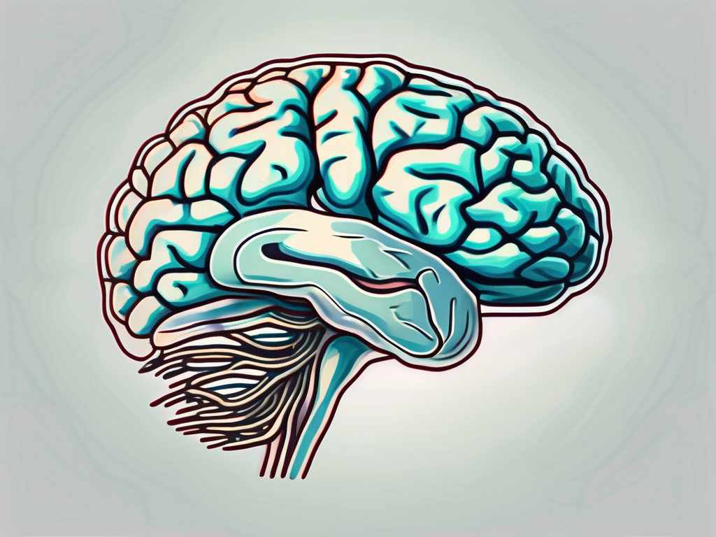
The abducens nerve, also known as cranial nerve VI, is a vital component of the complex network of nerves responsible for controlling eye movement. Derived from the head segment of the human body, the abducens nerve plays a crucial role in the coordination and alignment of the eyes, ensuring smooth and synchronized movement. Understanding the anatomy, function, and disorders related to this nerve can provide valuable insights into the intricate workings of the visual system.
Understanding the Abducens Nerve
The abducens nerve is a crucial component of the human visual system, playing a vital role in eye movement and coordination. This nerve, also known as the sixth cranial nerve, emerges from the brainstem, specifically the pons region, which is a part of the midbrain. Its anatomical pathway and functional significance make it an intriguing structure to explore.
Anatomy of the Abducens Nerve
The abducens nerve, being a motor nerve, controls the lateral rectus muscle, one of the six extraocular muscles responsible for moving the eye. This muscle’s primary function is to rotate the eye away from the midline of the face, allowing for horizontal gaze and facilitating binocular vision.
As the abducens nerve traverses its path, it enters the cavernous sinus, a space located within the skull, and eventually reaches the orbit, where it innervates the lateral rectus muscle. The intricate journey of this nerve through various anatomical structures highlights its significance in the overall functioning of the visual system.
Function of the Abducens Nerve
The primary function of the abducens nerve is to control the movement of the lateral rectus muscle, enabling the eye to move away from the midline of the face. This movement, known as abduction, is essential for horizontal gaze and facilitates binocular vision.
The synchronized coordination of the abducens nerve with other ocular motor nerves ensures accurate eye movement, contributing to depth perception and visual tracking. This coordination allows individuals to effortlessly shift their gaze from one point to another, following objects or scanning their surroundings.
Moreover, the abducens nerve’s role in eye movement is crucial for maintaining balance and spatial orientation. By controlling the lateral rectus muscle, this nerve helps individuals maintain stable vision while moving their heads or bodies.
Understanding the abducens nerve’s function is not only essential for comprehending the complexities of the visual system but also for diagnosing and treating various eye movement disorders. Dysfunction or damage to the abducens nerve can lead to a condition known as abducens nerve palsy, characterized by the inability to move the affected eye laterally.
In conclusion, the abducens nerve is a remarkable structure that plays a significant role in eye movement and coordination. Its anatomical pathway and functional significance highlight its importance in the overall functioning of the visual system. By controlling the lateral rectus muscle, the abducens nerve enables horizontal gaze, facilitates binocular vision, and contributes to depth perception and visual tracking. Further research and exploration of this nerve will undoubtedly enhance our understanding of the human visual system and potentially lead to advancements in the diagnosis and treatment of eye movement disorders.
Origin of the Abducens Nerve
The abducens nerve, also known as the sixth cranial nerve, has a fascinating origin during embryonic development. It arises from the basal plate of the neural tube, a structure that forms the central nervous system. Specifically, it originates from the ventral aspect of the medulla oblongata, which is a vital part of the brainstem responsible for various essential functions.
During the intricate process of embryogenesis, the abducens nerve gradually extends into the pons region, a bridge-like structure that connects the medulla oblongata to the midbrain. This extension is a remarkable example of the complexity and precision involved in the development of the nervous system.
Development of the Abducens Nerve
The development of the abducens nerve is a highly regulated process that involves the interaction of various molecular signals and genetic factors. These factors guide the growth and differentiation of neural cells, ensuring the proper formation of the nerve.
As the neural tube develops, specific regions give rise to different cranial nerves, including the abducens nerve. The basal plate, from which the abducens nerve originates, plays a crucial role in the development of motor neurons that control eye movement.
During embryogenesis, the abducens nerve undergoes a series of complex morphological changes, including axonal outgrowth and guidance. This process ensures that the nerve fibers reach their target muscles, allowing for precise control of eye movements.
Pathway of the Abducens Nerve
As the abducens nerve travels within the skull, it follows a specific pathway and shares the cavernous sinus with other vital structures. This sinus is a cavity located on each side of the sella turcica, a bony structure at the base of the skull.
Within the cavernous sinus, the abducens nerve coexists with important structures such as the carotid artery and other cranial nerves, including the oculomotor and trochlear nerves. This close proximity highlights the potential for the abducens nerve to be affected by various pathologies.
Disruptions along the pathway of the abducens nerve can occur due to various reasons, including vascular damage, inflammation, and tumors. These conditions can exert pressure on the nerve or interfere with its function, leading to impaired eye movement and, consequently, visual disturbances.
Understanding the intricate pathway of the abducens nerve and its vulnerability to potential pathologies is essential in diagnosing and treating conditions that affect eye movement. Medical professionals rely on detailed knowledge of the nerve’s anatomy and physiology to provide accurate assessments and develop appropriate treatment plans.
Head Segments and the Abducens Nerve
The abducens nerve, also known as cranial nerve VI, plays a crucial role in eye movement and coordination. It originates in the brainstem, specifically the pons region, highlighting its close association with this vital structure. The brainstem serves as a bridge between the brain and the spinal cord, housing various important nuclei and pathways responsible for essential bodily functions.
Any lesion or damage to the brainstem, whether due to trauma, tumors, or vascular events, can potentially affect the abducens nerve and compromise its function. The abducens nerve carries signals from the brain to the lateral rectus muscle, which is responsible for outward eye movement. If the abducens nerve is damaged, it can result in a condition called abducens nerve palsy, characterized by the inability to move the affected eye laterally.
It is essential to recognize the importance of maintaining the integrity of the brainstem to preserve the normal functioning of the abducens nerve and the visual system as a whole. The intricate network of nerves, muscles, and brain structures involved in eye movement requires precise coordination to ensure optimal visual acuity and binocular vision.
The Abducens Nerve and the Eye Muscles
The abducens nerve’s role in coordinating eye movements is closely tied to the functioning of the other extraocular muscles. These muscles work together to ensure precise eye alignment, allowing for smooth and coordinated movement in all directions. The six extraocular muscles, including the lateral rectus muscle innervated by the abducens nerve, work in harmony to control eye movements such as abduction, adduction, elevation, and depression.
Any disruption to the abducens nerve or the intricate balance of eye muscle coordination can lead to strabismus, commonly known as crossed eyes. Strabismus occurs when the eyes do not align properly, causing one eye to deviate from its normal position. This misalignment can significantly impact visual acuity and binocular vision, leading to difficulties in depth perception and visual integration.
Treatment options for strabismus vary depending on the underlying cause and severity of the condition. They may include corrective lenses, eye exercises, or surgical intervention to realign the eyes and restore proper eye muscle function.
In conclusion, the abducens nerve’s connection with the brainstem and its role in coordinating eye movements highlight its importance in maintaining optimal visual function. Understanding the intricate interplay between the abducens nerve, eye muscles, and other structures involved in eye movement is crucial in diagnosing and treating conditions that affect eye alignment and coordination.
Disorders Related to the Abducens Nerve
The abducens nerve, also known as the sixth cranial nerve, plays a crucial role in eye movement. When this nerve is affected by various disorders, specific symptoms may manifest. These symptoms can have a significant impact on a person’s vision and overall quality of life.
Symptoms of Abducens Nerve Disorders
One of the most common symptoms of abducens nerve disorders is double vision, also known as diplopia. This occurs when the affected eye is unable to align properly with the other eye, resulting in the perception of two images instead of one. Double vision can be extremely disorienting and can make it difficult to perform everyday tasks such as reading, driving, or even walking.
In addition to double vision, individuals with abducens nerve disorders may also experience difficulty moving the affected eye laterally. This means that they may have trouble looking towards the side of the affected eye, leading to a limited field of vision. This can be particularly challenging when trying to focus on objects or people located to the side.
Another noticeable symptom of abducens nerve disorders is a misalignment of the eyes, also known as strabismus. This occurs when the affected eye deviates from its normal position, causing one eye to appear misaligned or crossed. Strabismus can be not only a cosmetic concern but also a functional one, as it can affect depth perception and coordination.
If any of these symptoms occur, it is crucial to consult with a healthcare professional. Prompt evaluation and diagnosis are crucial for appropriate management and treatment of abducens nerve disorders. Only a qualified healthcare professional can accurately assess the underlying cause and recommend the most effective treatment plan.
Treatment and Management of Abducens Nerve Disorders
The treatment of abducens nerve disorders is highly dependent on the underlying cause. In some cases, conservative management may be effective in minimizing the visual symptoms associated with abducens nerve dysfunction. This can include the use of eye patches or prisms to help align the eyes and reduce double vision.
However, more severe cases of abducens nerve disorders may necessitate surgical intervention or other specialized treatments. Surgical options may involve adjusting the position of the eye muscles to improve alignment and reduce strabismus. These procedures are typically performed by ophthalmologists who specialize in eye muscle surgery.
It is important to note that the decision to pursue surgical intervention or other specialized treatments should only be made in consultation with a qualified healthcare professional. They will consider the individual’s specific condition, overall health, and personal preferences to determine the most appropriate course of action.
In conclusion, disorders related to the abducens nerve can have a significant impact on a person’s vision and overall well-being. Prompt evaluation, accurate diagnosis, and appropriate management are crucial for minimizing symptoms and improving quality of life. If you or someone you know is experiencing symptoms related to abducens nerve dysfunction, it is important to seek medical attention for proper evaluation and treatment.
The Role of the Abducens Nerve in Vision
The Abducens Nerve and Eye Movement
Eye movements are essential for visual perception, allowing us to explore our surroundings and focus on specific objects of interest. The abducens nerve plays a critical role in facilitating horizontal eye movements and ensuring accurate gaze control. Without the smooth functioning of the abducens nerve, our ability to track moving objects or maintain visual fixation would be significantly compromised.
The abducens nerve, also known as the sixth cranial nerve, originates from the pons, a region of the brainstem. It emerges from the brainstem and travels through the cavernous sinus, a cavity located behind the eyes. From there, it enters the orbit, the bony socket that houses the eyeball. Within the orbit, the abducens nerve innervates the lateral rectus muscle, which is responsible for outward eye movements.
When we look to the side, the abducens nerve sends signals to the lateral rectus muscle, causing it to contract and pull the eyeball outward. This coordinated movement allows both eyes to work together, ensuring that the visual field is covered efficiently. It is the abducens nerve that enables us to shift our gaze from one point to another smoothly, without jerky or uncontrolled movements.
The Abducens Nerve and Binocular Vision
Binocular vision, the ability to combine the visual inputs from both eyes into a single, coherent image, relies on the precise coordination of the abducens nerve and other ocular motor nerves. The integration of visual information from each eye enhances depth perception, enabling us to perceive three-dimensional space accurately. Any disruption to the abducens nerve can lead to binocular vision impairment, affecting daily activities such as reading, driving, and sports.
When both eyes are aligned properly, the abducens nerve ensures that the visual information received by each eye is fused together in the brain, creating a unified and three-dimensional perception of the world. This fusion of visual inputs is crucial for tasks that require depth perception, such as judging distances, catching a ball, or navigating through a crowded environment.
In cases where the abducens nerve is not functioning correctly, binocular vision problems can arise. This can manifest as double vision, where two slightly different images are perceived simultaneously, or as a misalignment of the eyes, known as strabismus. Strabismus can lead to amblyopia, commonly known as lazy eye, where the brain suppresses the visual input from one eye to avoid confusion. Early detection and treatment of abducens nerve disorders are essential to prevent long-term vision problems and ensure optimal visual development.
In conclusion, the abducens nerve, derived from the head segment of the human body, plays a crucial role in eye movement coordination. Understanding the intricate anatomy, function, and disorders related to this nerve provides valuable insights into the complex dynamics of the visual system. If you experience any visual symptoms or suspect abducens nerve dysfunction, it is essential to consult with a healthcare professional for appropriate evaluation and management. Appreciating the delicate interplay between the abducens nerve and the visual system allows us to appreciate the remarkable complexity of our ability to perceive the world around us.

Leave a Reply