The human eye is a complex and remarkable organ, responsible for our vision and allowing us to perceive the world around us. To fully understand the intricate workings of the eye, it is essential to have a grasp of its anatomy and the role of its various components. One aspect that deserves attention is the connection between the abducens nerve (CN VI) and a specific extrinsic eye muscle. In this article, we will explore the anatomy of the eye, the function of the abducens nerve, and the significance of its innervation on eye movement.
Understanding the Anatomy of the Eye
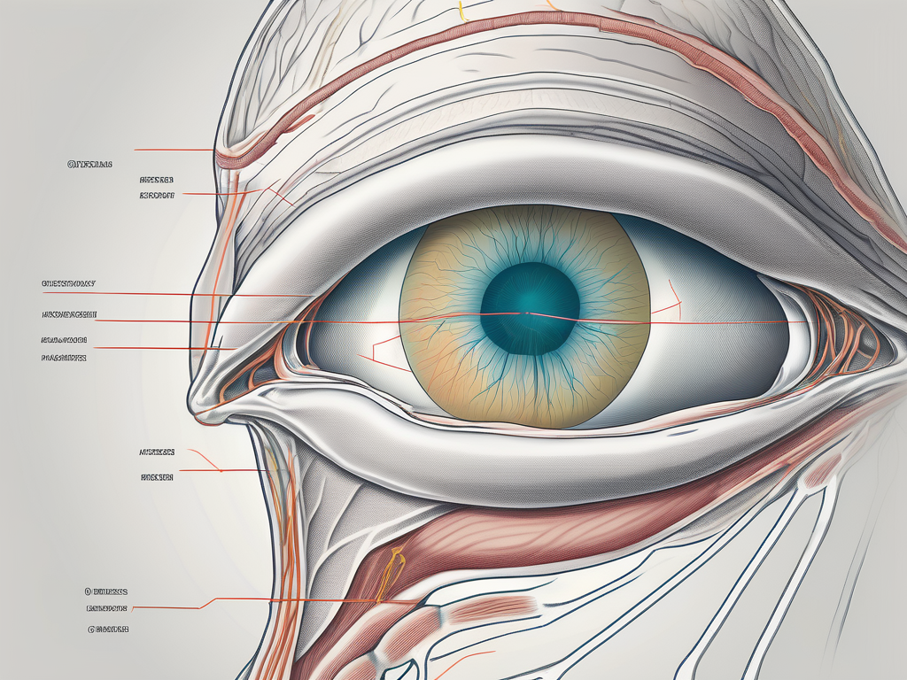
The eye is a sensory organ situated within the orbit of the skull. Its primary function is to receive visual stimuli and transmit them to the brain for interpretation. The eye is composed of several components, including the cornea, iris, lens, retina, and optic nerve. These structures work in harmony to ensure the accurate capture and processing of visual information.
The cornea, a transparent dome-shaped structure, is the outermost layer of the eye. It acts as a protective barrier, shielding the delicate inner structures from external elements. The cornea also plays a crucial role in focusing light onto the retina, helping to create clear and sharp images.
Located behind the cornea is the iris, which is the colored part of the eye. The iris controls the amount of light entering the eye by adjusting the size of the pupil. In bright conditions, the iris contracts, making the pupil smaller to reduce the amount of light. Conversely, in dim lighting, the iris expands, allowing more light to enter through a larger pupil.
The lens, situated behind the iris, further focuses the incoming light onto the retina. It is a flexible and transparent structure that can change its shape to adjust the focus, allowing us to see objects at different distances. This process, known as accommodation, is crucial for clear vision at various distances.
The retina, located at the back of the eye, is a thin layer of tissue composed of specialized cells called photoreceptors. These photoreceptors, known as rods and cones, convert light into electrical signals that can be interpreted by the brain. Rods are responsible for vision in low-light conditions, while cones are responsible for color vision and visual acuity.
The optic nerve, often referred to as the “information highway” of the eye, carries the electrical signals generated by the retina to the brain. It is composed of millions of nerve fibers that transmit visual information to the visual cortex, where it is processed and interpreted, allowing us to perceive the world around us.
The Role of Extrinsic Eye Muscles
While the internal structures of the eye are vital for vision, there are also external factors that contribute to its functionality. Extrinsic eye muscles, a group of six muscles located outside the eye, are responsible for controlling its movement. These muscles work synergistically to allow smooth, coordinated eye movements in various directions.
The superior rectus muscle, located above the eye, is responsible for upward eye movement. It helps us look up at objects or raise our gaze to the sky. On the other hand, the inferior rectus muscle, situated below the eye, facilitates downward eye movement, allowing us to look down or lower our gaze.
The medial rectus muscle, positioned towards the nose, enables inward eye movement, allowing us to focus on objects that are closer to us. Conversely, the lateral rectus muscle, located towards the temple, facilitates outward eye movement, enabling us to look towards the sides.
The superior oblique muscle, originating from the back of the eye socket, plays a crucial role in downward and inward eye movement. It helps us look down and towards the nose. Lastly, the inferior oblique muscle, originating from the front of the eye socket, contributes to upward and outward eye movement, allowing us to look up and towards the temple.
The Function of the Abducens Nerve (CN VI)
Among the cranial nerves involved in eye movement, the abducens nerve (CN VI) plays a crucial role. This nerve, originating from the brainstem, serves as the primary source of motor innervation for one specific extrinsic eye muscle. By controlling the contraction and relaxation of this muscle, the abducens nerve facilitates lateral eye movement, specifically abduction.
Abduction refers to the movement of the eye away from the midline of the face, towards the temple. This movement allows us to look sideways, scanning our surroundings or shifting our gaze to objects located to the sides. The abducens nerve ensures that this movement is smooth and coordinated, allowing us to navigate our environment effectively.
The Connection between the Abducens Nerve and Eye Movement
The ability to move our eyes freely in different directions is essential for optimal visual function. The abducens nerve’s intricate connection to an extrinsic eye muscle ensures the smooth execution of specific eye movements.
The abducens nerve, also known as the sixth cranial nerve, is responsible for controlling the movement of the eye. It is one of the twelve pairs of cranial nerves that originate in the brainstem and extend to various parts of the head and neck. Specifically, the abducens nerve innervates the lateral rectus muscle, which is one of the six extraocular muscles that control eye movement.
How the Abducens Nerve Controls Eye Movement
Through its innervation of the lateral rectus muscle, the abducens nerve plays a pivotal role in horizontal eye movement. When the abducens nerve is stimulated, it signals the lateral rectus muscle to contract, causing the eye to move away from the midline of the face. This lateral movement is fundamental for activities such as scanning an environment or tracking a moving object.
Imagine you are walking down a busy street, trying to take in all the sights and sounds around you. Your abducens nerve is hard at work, coordinating the movement of your eyes as they shift from one point of interest to another. Without the proper functioning of the abducens nerve, your ability to explore your surroundings would be severely limited.
The Impact of Abducens Nerve Damage on Eye Function
Unfortunately, the abducens nerve is susceptible to damage and dysfunction. When the abducens nerve is impaired, it can lead to a condition known as abducens nerve palsy. This condition results in the inability to fully abduct the affected eye, causing double vision and limited eye movement towards the affected side.
Abducens nerve palsy can have various causes, including trauma, infections, tumors, or underlying medical conditions such as diabetes or multiple sclerosis. The severity of the symptoms may vary depending on the extent of the nerve damage.
If you experience any concerning symptoms or suspect abducens nerve involvement, it is crucial to consult with a medical professional for a proper diagnosis and appropriate management. Treatment options for abducens nerve palsy may include medications, eye exercises, or in some cases, surgical intervention.
Research and advancements in the field of neurology continue to shed light on the intricate connection between the abducens nerve and eye movement. Understanding the complexities of this relationship not only helps in diagnosing and treating conditions related to the abducens nerve but also contributes to our overall knowledge of the human visual system.
Identifying the Specific Eye Muscle Innervated by the Abducens Nerve
Among the extrinsic eye muscles, the lateral rectus muscle is the sole muscle innervated by the abducens nerve. Understanding the anatomy and function of the lateral rectus muscle enhances our comprehension of the abducens nerve’s role in eye movement.
The Lateral Rectus Muscle: An Overview
The lateral rectus muscle is one of the six extrinsic eye muscles responsible for eye movement. Located on the lateral side of the eye, it contracts to move the eye laterally away from the midline. Its antagonist muscle, the medial rectus, performs the opposite action, medially moving the eye.
Let’s delve deeper into the structure and function of the lateral rectus muscle. This muscle originates from the common tendinous ring, also known as the annulus of Zinn, which surrounds the optic nerve. From there, it extends laterally towards the outer edge of the eye.
The lateral rectus muscle is composed of striated muscle fibers, which are under voluntary control. These fibers are innervated by the abducens nerve, also known as cranial nerve VI (CN VI). The abducens nerve originates from the pons, a region of the brainstem, and travels through the cavernous sinus before reaching the lateral rectus muscle.
When the abducens nerve sends motor impulses to the lateral rectus muscle, it contracts, causing the eye to move laterally. This movement is essential for horizontal gaze and allows us to shift our focus to objects located to the sides.
The Relationship between the Lateral Rectus Muscle and the Abducens Nerve
The abducens nerve (CN VI) innervates the lateral rectus muscle, providing it with the necessary motor impulses for contraction. This unique connection ensures the precise control of lateral eye movement, allowing us to shift our gaze to the sides.
Let’s explore the intricate relationship between the abducens nerve and the lateral rectus muscle in more detail. The abducens nerve arises from the abducens nucleus, which is located in the pons of the brainstem. From there, it travels through the cavernous sinus, a cavity located in the skull, before reaching the lateral rectus muscle.
Upon reaching the lateral rectus muscle, the abducens nerve branches out into multiple motor fibers, which innervate the muscle fibers of the lateral rectus. These motor fibers transmit electrical signals from the brain to the muscle, triggering its contraction.
The precise coordination between the abducens nerve and the lateral rectus muscle is crucial for smooth and accurate eye movements. Any disruption in this connection can lead to impaired lateral eye movement, resulting in difficulties in focusing on objects located to the sides.
It is worth noting that the lateral rectus muscle works in conjunction with other extrinsic eye muscles to ensure coordinated eye movements. For example, when we shift our gaze to the right, the right lateral rectus muscle contracts, while the left medial rectus muscle contracts simultaneously to medially move the left eye.
In conclusion, the abducens nerve plays a vital role in eye movement by innervating the lateral rectus muscle. This connection allows for precise control of lateral eye movement, enabling us to shift our gaze to the sides effortlessly. Understanding the intricate relationship between the abducens nerve and the lateral rectus muscle enhances our knowledge of the complex mechanisms involved in eye movement.
The Clinical Significance of the Abducens Nerve and Eye Muscles
The abducens nerve and the lateral rectus muscle have a crucial relationship that is essential for recognizing and managing various eye disorders. Understanding this relationship is vital for comprehending the functionality of the eye and the potential impact of abducens nerve dysfunction.
The abducens nerve is responsible for the innervation of the lateral rectus muscle, which is one of the six extraocular muscles that control eye movement. This muscle is specifically responsible for moving the eye laterally, allowing for horizontal gaze and facilitating binocular vision.
Common Disorders Involving the Abducens Nerve
Abducens nerve palsy is one of the most common disorders affecting the abducens nerve. It occurs when there is damage or dysfunction of the nerve, leading to a range of visual disturbances. This condition can result from various causes, including trauma, infections, vascular conditions, or other underlying systemic diseases.
Individuals with abducens nerve palsy often experience a decreased ability to move their affected eye laterally. This limitation can lead to double vision (diplopia) and difficulty in focusing on objects located to the side. In some cases, the affected eye may also deviate inward, causing an inward squint (esotropia).
It is important to note that abducens nerve palsy can occur unilaterally (affecting one eye) or bilaterally (affecting both eyes). Bilateral abducens nerve palsy is a more severe condition and can significantly impact a person’s ability to coordinate eye movements.
Treatment and Management of Abducens Nerve Disorders
The treatment and management options for abducens nerve disorders depend on the underlying cause and severity of the condition. In some cases, conservative measures such as eye exercises and the use of prisms may help improve the patient’s symptoms.
Eye exercises can help strengthen the affected eye muscles and improve their coordination. These exercises often involve focusing on specific targets and moving the eyes in different directions to enhance muscle control and flexibility.
Prisms are optical devices that can be incorporated into eyeglasses to help align the visual images from both eyes, reducing double vision. By redirecting light rays, prisms can compensate for the misalignment caused by abducens nerve dysfunction.
In more severe cases or when conservative measures are ineffective, surgical intervention may be necessary. Surgical options for abducens nerve disorders include procedures to correct muscle imbalances or to reposition the affected eye muscles for improved alignment.
It is crucial to consult with an experienced ophthalmologist or neurologist to determine the most appropriate treatment plan for your specific condition. These specialists can conduct a thorough evaluation, including a comprehensive eye examination and diagnostic tests, to identify the underlying cause of the abducens nerve disorder and develop an individualized treatment approach.
Regular follow-up appointments with your healthcare professional are essential to monitor the progress of the treatment and make any necessary adjustments. They can also provide guidance on managing any persistent symptoms or addressing any potential complications that may arise.
In conclusion, the abducens nerve (CN VI) plays a critical role in eye movement, specifically the contraction of the lateral rectus muscle. Understanding the intricate connection between the abducens nerve and the lateral rectus muscle allows us to comprehensively comprehend the eye’s functionality and recognize the potential impact of abducens nerve dysfunction.
If you experience any concerning symptoms related to eye movement or suspect abducens nerve involvement, it is crucial to seek medical advice and consult with a healthcare professional specialized in ophthalmology or neurology. Early diagnosis and appropriate management can significantly improve outcomes and enhance the quality of life for individuals with abducens nerve disorders.
Leave a Reply