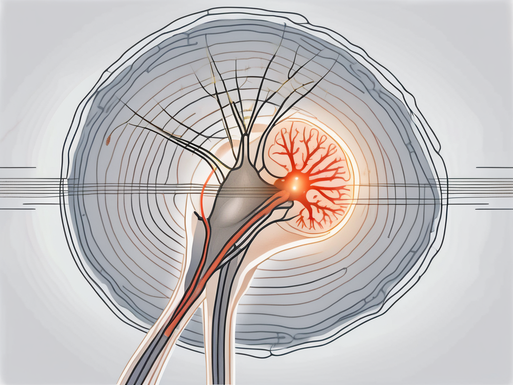
The abducens nerve, also known as cranial nerve VI, is responsible for the innervation of a specific muscle called the lateral rectus muscle. Understanding the function and anatomy of the abducens nerve, as well as its relationship with the lateral rectus muscle, is crucial to comprehend its significance in eye movement. Additionally, it is essential to recognize the medical conditions related to the abducens nerve and the recent advancements in its research.
Understanding the Abducens Nerve
The abducens nerve, also known as cranial nerve VI, is one of the twelve cranial nerves originating in the brainstem. It plays a vital role in controlling the movement of the eye, specifically in the outward direction. Without the proper functioning of the abducens nerve, coordinated eye movement and maintaining binocular vision would be challenging.
The abducens nerve has a fascinating anatomy. It is a motor nerve that arises from the abducens nucleus located in the pons region of the brainstem. From there, it emerges from the brain and travels through a bony canal called the cavernous sinus. This intricate pathway ensures that the nerve reaches its destination – the orbit of the eye, where it innervates the lateral rectus muscle.
The abducens nerve carries signals from the brain to the lateral rectus muscle, instructing it to contract and move the eye laterally. This lateral movement allows the eyes to gaze away from the midline, such as when looking towards the temple or the periphery of our visual field.
Anatomy of the Abducens Nerve
The abducens nerve originates from the abducens nucleus, which is located in the pons region of the brainstem. This nucleus contains the cell bodies of the nerve fibers that make up the abducens nerve. From the nucleus, the nerve fibers exit the brainstem and travel through the cavernous sinus, a complex network of veins and nerves located on either side of the sella turcica, a bony structure in the skull.
As the abducens nerve traverses the cavernous sinus, it passes through several important structures, including the internal carotid artery and the oculomotor and trochlear nerves. These structures are crucial for maintaining the blood supply to the brain and coordinating eye movements.
After leaving the cavernous sinus, the abducens nerve enters the orbit of the eye through the superior orbital fissure, a narrow opening in the skull. Once inside the orbit, the nerve innervates the lateral rectus muscle, which is responsible for the abduction of the eye.
Function of the Abducens Nerve
The primary function of the abducens nerve is to control the movement of the lateral rectus muscle. When the abducens nerve is activated, it sends signals to the lateral rectus muscle, causing it to contract. This contraction leads to the abduction of the eye, allowing it to move away from the midline.
Proper functioning of the abducens nerve is crucial for maintaining binocular vision and coordinating eye movements. When both eyes work together, they provide depth perception and a wider field of view. Impairment or damage to the abducens nerve can lead to various visual disturbances and eye movement disorders.
Conditions that affect the abducens nerve can result in a range of symptoms, including double vision (diplopia), difficulty moving the eye laterally, and misalignment of the eyes (strabismus). These symptoms can significantly impact a person’s ability to perform everyday tasks that require coordinated eye movements, such as reading, driving, and playing sports.
Diagnosing and treating abducens nerve disorders often involve a comprehensive evaluation by a neurologist or ophthalmologist. They may conduct a thorough medical history review, physical examination, and specialized tests, such as eye movement recordings and imaging studies, to determine the underlying cause of the problem.
Treatment options for abducens nerve disorders depend on the specific condition and its severity. In some cases, conservative measures, such as eye exercises or prism glasses, may be sufficient to manage the symptoms. However, more severe cases may require surgical intervention to correct the underlying issue or alleviate pressure on the nerve.
Overall, the abducens nerve plays a crucial role in eye movement and maintaining binocular vision. Understanding its anatomy and function can help healthcare professionals diagnose and manage disorders affecting this vital cranial nerve.
The Muscle Innervated by the Abducens Nerve
Identifying the Lateral Rectus Muscle
The lateral rectus muscle is one of the six extraocular muscles that control eye movement. It is located on the outer side of each eye and is responsible for moving the eye laterally. The lateral rectus muscle works in opposition to the medial rectus muscle, which brings the eye back towards the midline.
When the abducens nerve sends signals to the lateral rectus muscle, it contracts, causing the eye to move away from the midline. This movement is essential for horizontal gaze and allows us to track objects in our peripheral vision.
The lateral rectus muscle, with its unique positioning on the outer side of each eye, plays a crucial role in maintaining eye alignment and facilitating coordinated eye movements. It is responsible for the lateral movement of the eye, allowing us to explore our visual surroundings with ease.
Imagine standing in a bustling city street, surrounded by a myriad of sights and sounds. As you turn your head to the left, your lateral rectus muscles come into action, pulling your eyes towards the side of your head. This movement enables you to scan the environment, taking in the vibrant colors, the movement of people, and the architecture that surrounds you.
Furthermore, the lateral rectus muscle works in harmony with the other extraocular muscles, ensuring smooth and accurate eye movements in different directions. Together, these muscles allow us to shift our gaze effortlessly, whether it’s following the flight of a butterfly or tracking a speeding car.
Role of the Lateral Rectus Muscle in Eye Movement
The lateral rectus muscle plays a critical role in maintaining eye alignment and facilitating coordinated eye movements. It works together with the other extraocular muscles to ensure smooth and accurate eye movements in different directions.
When the lateral rectus muscle contracts, it pulls the eye towards the side of the head, allowing for horizontal movement. This action is particularly important when looking towards objects located away from the midline, such as when scanning the environment or following the movements of a moving object.
Imagine sitting in a park on a sunny day, watching a flock of birds flying overhead. As the birds soar across the sky, your lateral rectus muscles come into play, allowing your eyes to smoothly track their flight. The lateral rectus muscle ensures that your gaze follows the birds’ movement, providing you with a captivating visual experience.
Moreover, the lateral rectus muscle’s role in eye movement extends beyond simple tracking. It also contributes to our ability to maintain binocular vision, which is crucial for depth perception. By coordinating with the other eye muscles, the lateral rectus muscle helps align both eyes, allowing us to perceive the world in three dimensions.
Next time you find yourself marveling at a breathtaking landscape or engrossed in a thrilling movie, remember to thank your lateral rectus muscles for their tireless work. Without them, our visual experiences would lack the richness and depth that make life so vibrant.
The Relationship between the Abducens Nerve and Lateral Rectus Muscle
The abducens nerve and the lateral rectus muscle play a crucial role in eye movement and alignment. Understanding how these two components work together is essential in comprehending the complex mechanics of the human visual system.
How the Abducens Nerve Controls the Lateral Rectus Muscle
The abducens nerve, also known as the sixth cranial nerve, is responsible for transmitting signals from the brain to the lateral rectus muscle. This muscle is one of the six extraocular muscles that control eye movement. Specifically, the abducens nerve instructs the lateral rectus muscle to contract and move the eye laterally.
When the abducens nerve is functioning properly, it ensures the precise control of eye movement, allowing for the appropriate alignment of both eyes. The impulses generated in the abducens nucleus, located in the pons of the brainstem, travel along the abducens nerve fibers until they reach the lateral rectus muscle.
Upon reaching the lateral rectus muscle, these impulses trigger the release of neurotransmitters, which initiate muscle contraction. The contraction of the lateral rectus muscle results in the desired eye movement, allowing the eye to move laterally and maintain proper alignment with the other eye.
Implications of Damage to the Abducens Nerve
Damage to the abducens nerve can have significant implications on eye movement and alignment. One common condition associated with abducens nerve damage is abducens nerve palsy. This condition is characterized by the weakness or paralysis of the lateral rectus muscle.
When the abducens nerve is damaged, the affected eye’s ability to move laterally is hampered, resulting in a limited range of motion and potential misalignment with the other eye. This misalignment, known as strabismus, can lead to double vision and difficulty with horizontal eye movements.
If you experience any of these symptoms, it is crucial to consult with a healthcare professional for an accurate diagnosis and appropriate treatment. Treatment options for abducens nerve palsy may include eye exercises, prism glasses, or in severe cases, surgical intervention to correct the misalignment and restore proper eye movement.
Understanding the intricate relationship between the abducens nerve and the lateral rectus muscle is essential in comprehending the complexities of the human visual system. Further research and advancements in medical science continue to shed light on these mechanisms, paving the way for improved diagnostic techniques and treatment options for individuals affected by abducens nerve-related conditions.
Medical Conditions Related to the Abducens Nerve
The abducens nerve, also known as the sixth cranial nerve, plays a crucial role in controlling the movement of the eye. When this nerve is affected by certain medical conditions, it can lead to a condition known as abducens nerve palsy. Abducens nerve palsy can occur due to various underlying causes, including head trauma, infection, tumors, or neurological conditions such as multiple sclerosis.
Regardless of the cause, the common symptoms of abducens nerve palsy include reduced or absent lateral eye movement, eye misalignment, and diplopia (double vision). These symptoms can significantly impact a person’s quality of life and daily activities, making it essential to seek medical attention if they are experienced.
If you experience any of these symptoms, it is essential to seek medical attention. A healthcare professional can perform a comprehensive assessment, including a detailed medical history, physical examination, and possibly specialized tests, to determine the underlying cause of your symptoms. This thorough evaluation is crucial in order to provide an accurate diagnosis and develop an appropriate treatment plan.
Symptoms of Abducens Nerve Palsy
Abducens nerve palsy can manifest in various ways, depending on the severity and underlying cause of the condition. In addition to the common symptoms mentioned earlier, individuals with abducens nerve palsy may also experience eye fatigue, headaches, and difficulties with depth perception.
The reduced or absent lateral eye movement can make it challenging to focus on objects located to the side, affecting tasks such as reading, driving, or participating in sports. Eye misalignment can lead to a noticeable cosmetic change, causing self-consciousness and affecting self-esteem.
Diplopia, or double vision, occurs when the eyes are unable to align properly, resulting in the perception of two images instead of one. This can cause significant visual discomfort and make it difficult to perform everyday activities that require clear vision, such as reading, watching television, or using a computer.
It is important to note that the severity of symptoms may vary from person to person, depending on the extent of nerve damage and the underlying cause. Therefore, a comprehensive evaluation by a healthcare professional is crucial to determine the appropriate course of treatment.
Treatment Options for Abducens Nerve Disorders
Treatment options for abducens nerve disorders depend on the underlying cause and severity of the condition. In some cases, conservative treatments such as eye patching, prism glasses, or vision therapy may be recommended to improve eye alignment and reduce double vision.
Eye patching involves covering the stronger eye to encourage the weaker eye to strengthen and regain its normal function. Prism glasses work by redirecting light, helping to align the eyes and reduce double vision. Vision therapy is a specialized form of therapy that aims to improve eye coordination and strengthen the eye muscles through various exercises and techniques.
If the abducens nerve palsy is caused by an underlying medical condition, such as a tumor or infection, specific treatments targeting the root cause may be necessary. These may involve surgical interventions, medications, or other specialized therapies. It is essential to consult with a healthcare professional who can provide individualized treatment recommendations based on your specific condition.
It is worth noting that early intervention and prompt treatment can significantly improve the prognosis and minimize the long-term effects of abducens nerve disorders. Therefore, if you suspect any issues with your eye movement or experience any of the symptoms mentioned, it is crucial to seek medical attention as soon as possible.
Recent Research on the Abducens Nerve and Lateral Rectus Muscle
Advances in Understanding the Abducens Nerve
In recent years, researchers have made significant strides in understanding the complex nature of the abducens nerve and its role in coordinating eye movements. The abducens nerve, also known as the sixth cranial nerve, is responsible for the movement of the lateral rectus muscle, which controls the outward movement of the eye.
Advances in neuroimaging techniques, such as magnetic resonance imaging (MRI) and functional MRI (fMRI), have allowed scientists to visualize the structure and activity of the abducens nerve in greater detail. Through these imaging techniques, researchers have been able to map the pathway of the abducens nerve from its origin in the brainstem to its innervation of the lateral rectus muscle.
These advancements have not only improved our understanding of normal abducens nerve function, but they have also aided in identifying abnormalities and potential treatment targets in patients with abducens nerve disorders. Conditions such as abducens nerve palsy, which is characterized by the inability to move the affected eye laterally, can now be better diagnosed and managed.
Future Directions in Abducens Nerve Research
Looking ahead, researchers are focusing on further unraveling the intricate connections between the abducens nerve, the brainstem, and other regions involved in eye movement and coordination. Understanding how the abducens nerve interacts with other cranial nerves, such as the oculomotor nerve and the trochlear nerve, will provide a more comprehensive understanding of eye movement control.
Investigating the molecular and cellular mechanisms underlying abducens nerve function may provide valuable insights into potential therapeutic approaches for eye movement disorders. Researchers are studying the genes and proteins involved in the development and maintenance of the abducens nerve, as well as the signaling pathways that regulate its activity.
Furthermore, the integration of emerging technologies, such as optogenetics and gene therapy, holds promise for developing innovative treatments that target specific components of the abducens nerve and related structures. Optogenetics, for example, involves the use of light-sensitive proteins to control the activity of neurons, offering a potential avenue for restoring normal abducens nerve function in patients with movement disorders.
Overall, the continued exploration of the abducens nerve and its relationship with the lateral rectus muscle paves the way for advancements in understanding eye movement disorders and improving patient care. Collaborative efforts between researchers, clinicians, and other healthcare professionals will undoubtedly lead to further breakthroughs in this fascinating field of study.

Leave a Reply