Everything You Wanted to Know About Abducens Nerve!
What is Abducens Nerve?
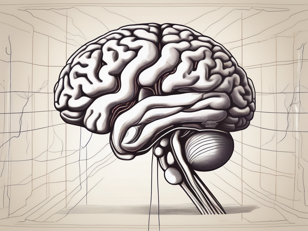
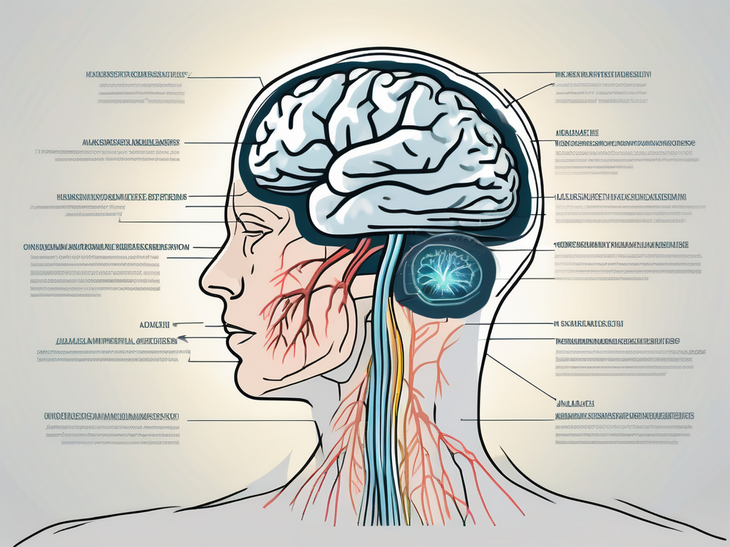
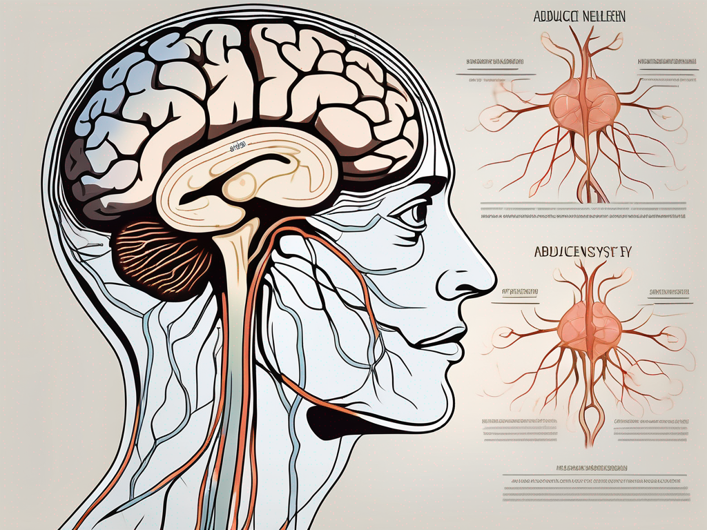
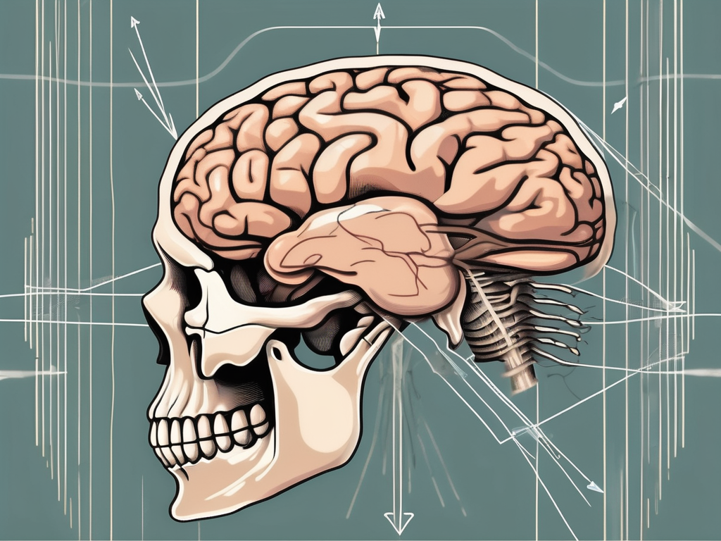
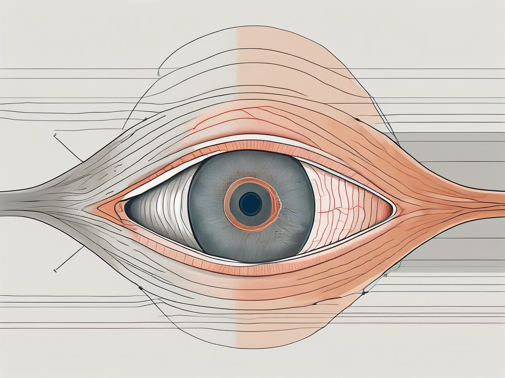
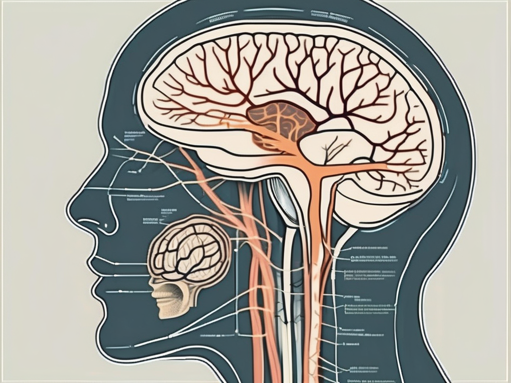
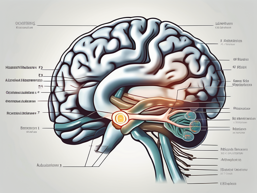
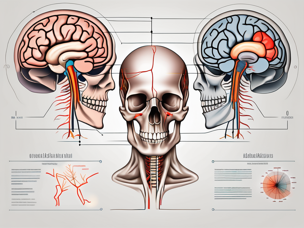
The abducens nerve, also known as cranial nerve VI, plays a crucial role in the control of eye movement.
Understanding the anatomy and function of this nerve is essential for physicians, researchers, and individuals interested in the complex workings of the human body.
-
what is the function of the abducens nerve
Discover the crucial role of the abducens nerve in controlling eye movement and maintaining visual coordination.
-
which nerve is the abducens nerve) palsy
Discover the ins and outs of abducens nerve palsy in this comprehensive article.
-
the abducens nerve serves what function?
Discover the crucial role of the abducens nerve in controlling eye movement and maintaining visual coordination.
-
which extrinsic eye muscle is innervated by the abducens nerve (cn vi)?
Uncover the fascinating connection between the abducens nerve (CN VI) and the extrinsic eye muscle it innervates in this insightful article.
-
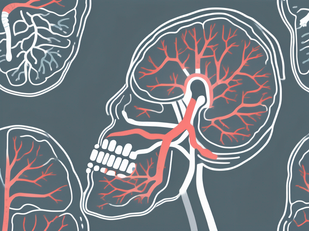
abducens nerve travels through what sinus
Discover the fascinating journey of the abducens nerve as it navigates through the intricate network of sinuses in the human skull.
-
what causes abducens nerve palsy
Explore the underlying factors and potential causes of abducens nerve palsy in this insightful article.
-
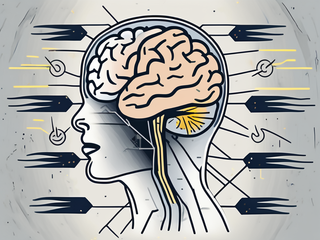
what happens if the abducens nerve is damaged
Discover the potential consequences of damage to the abducens nerve in this insightful article.
-
what is the innervation of the abducens nerve
Uncover the intricate web of nerve connections in the human body as you delve into the fascinating world of the abducens nerve innervation.
-
what does the abducens nerve serve
Discover the fascinating role of the abducens nerve in eye movement and its connection to various neurological conditions.
-
what is abducens nerve pair
Discover the intricate functions of the abducens nerve pair in this insightful article.