The abducens nerve, also known as cranial nerve VI, is a crucial component of the nervous system. Understanding its origin, function, anatomy, and associated disorders is essential for gaining a comprehensive understanding of this intricate neural pathway.
Understanding the Abducens Nerve
As one of the twelve cranial nerves, the abducens nerve plays a pivotal role in eye movement and coordination. Although it may be considered one of the smaller nerves, its impact on visual perception and ocular control is significant.
Definition and Function of the Abducens Nerve
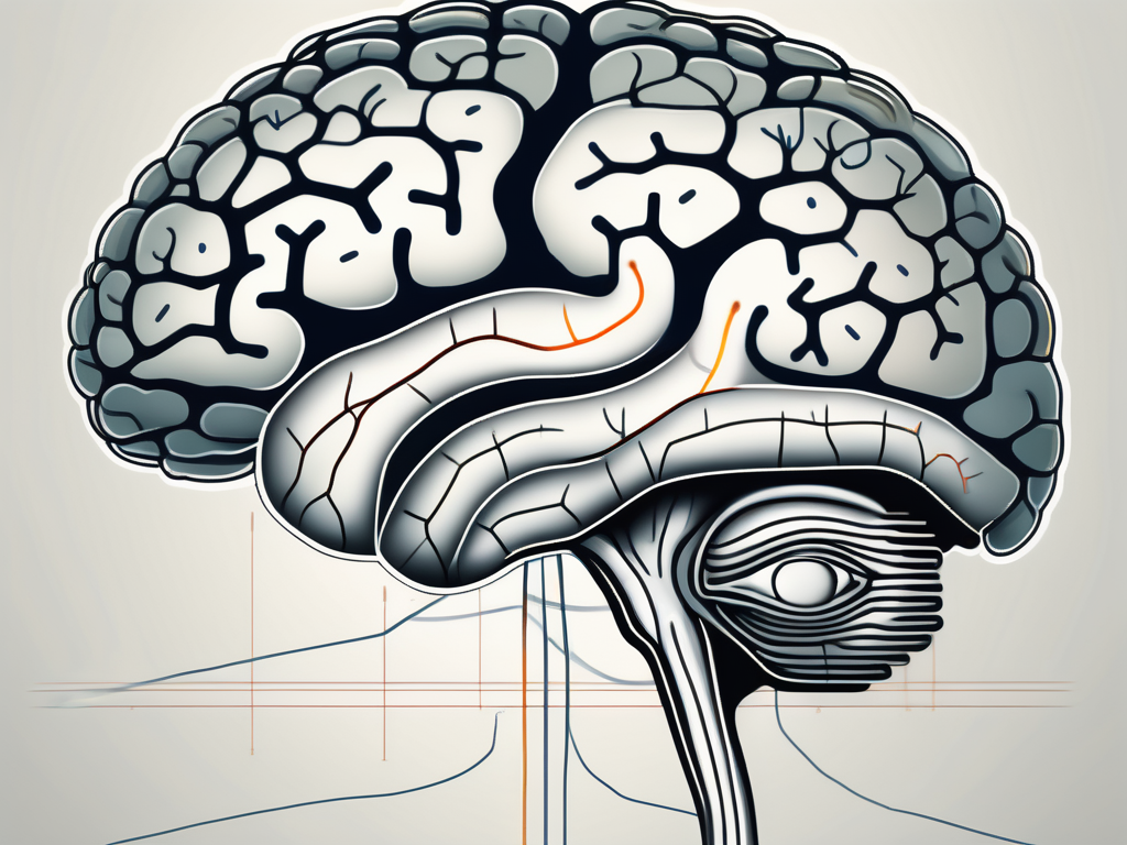
The abducens nerve originates in the brainstem, specifically the pons. It innervates the lateral rectus muscle, which is responsible for the outward movement of the eye, a movement known as abduction. This nerve’s name, “abducens,” is derived from its unique role in this particular eye movement, enabling us to look sideways.
The abducens nerve is part of the somatic motor division of the peripheral nervous system. It carries signals from the brain to the lateral rectus muscle, instructing it to contract and move the eye laterally. This movement is crucial for our ability to explore our environment and shift our focus efficiently.
Interestingly, the abducens nerve is the only cranial nerve that emerges from the brainstem in a ventral direction. This unique anatomical feature allows it to reach the lateral rectus muscle, which is located on the outer side of the eye, and control its movement effectively.
The Role of the Abducens Nerve in Eye Movement
Eye movements are complex, requiring precise coordination of multiple nerves and muscles. The abducens nerve’s primary function is to control the lateral rectus muscle, enabling the eye to gaze away from the midline. Consequently, it collaborates with other cranial nerves to facilitate coordinated eye movements, allowing us to explore our surroundings and shift our focus efficiently.
When the abducens nerve is functioning properly, it ensures that both eyes move in synchrony, allowing us to maintain binocular vision. This coordination is crucial for depth perception and accurate visual processing. However, when there is a dysfunction or damage to the abducens nerve, it can lead to a condition called abducens nerve palsy.
Abducens nerve palsy is characterized by the inability to move the affected eye laterally, resulting in a condition known as strabismus. Strabismus, commonly referred to as crossed eyes, can cause double vision and affect the individual’s ability to focus on objects. Treatment for abducens nerve palsy may involve eye exercises, patching, or in severe cases, surgical intervention.
In addition to its role in eye movement, the abducens nerve also plays a role in maintaining balance and coordination. It receives sensory information from the vestibular system, which is responsible for detecting changes in head position and movement. This input allows the brain to make adjustments to eye position and movement, ensuring that our visual perception remains stable even during rapid head movements.
Overall, the abducens nerve is a vital component of our visual system, enabling us to explore our surroundings, shift our focus, and maintain binocular vision. Its intricate connection with other cranial nerves and muscles highlights the complexity of eye movement and the remarkable coordination required for our visual perception to function optimally.
Anatomy of the Abducens Nerve
Examining the anatomy of the abducens nerve provides valuable insight into its location, pathway, and relationships with other cranial nerves.
The abducens nerve, also known as the sixth cranial nerve or cranial nerve VI, plays a crucial role in eye movement. It is responsible for controlling the lateral rectus muscle, which is responsible for abducting or moving the eye laterally away from the midline.
Location and Pathway of the Abducens Nerve
The abducens nerve emerges from the pons region of the brainstem, near the intersection of the medulla and the midbrain. This region is often referred to as the pontomedullary junction. From its origin, the nerve takes a long and intricate pathway through the skull.
After leaving the brainstem, the abducens nerve enters the cavernous sinus, a dural venous sinus located within the skull. The cavernous sinus is a complex network of veins that houses several important structures, including the internal carotid artery, oculomotor nerve (cranial nerve III), trochlear nerve (cranial nerve IV), and ophthalmic division of the trigeminal nerve (cranial nerve V1).
Within the cavernous sinus, the abducens nerve travels alongside the internal carotid artery, which supplies blood to the brain. This close proximity to the artery exposes the abducens nerve to potential compression or damage in cases of vascular abnormalities or tumors.
Continuing its journey, the abducens nerve exits the cavernous sinus through the superior orbital fissure, a narrow opening located in the sphenoid bone. It then enters the orbit, the bony socket that houses the eye.
Finally, the abducens nerve reaches its destination, the lateral rectus muscle. This muscle is one of the six extraocular muscles responsible for eye movements. When the abducens nerve stimulates the lateral rectus muscle, it causes the eye to move laterally, allowing for horizontal gaze and abduction.
Connection to Other Cranial Nerves
The abducens nerve maintains close associations with other cranial nerves, particularly the oculomotor (cranial nerve III) and trochlear (cranial nerve IV) nerves. These three nerves work together to coordinate the movements of the eyes, allowing our visual system to function harmoniously.
The oculomotor nerve innervates several extraocular muscles, including the superior rectus, inferior rectus, and inferior oblique muscles. It is responsible for upward and downward eye movements, as well as inward rotation of the eye. The abducens nerve complements the oculomotor nerve’s actions by controlling the lateral rectus muscle, which moves the eye laterally.
The trochlear nerve, on the other hand, innervates the superior oblique muscle. This muscle plays a crucial role in downward and inward eye movements. The coordinated actions of the abducens, oculomotor, and trochlear nerves allow for smooth and precise eye movements in various directions.
Any disruption or dysfunction within this network of cranial nerves may lead to significant visual impairments. Conditions such as abducens nerve palsy, oculomotor nerve palsy, or trochlear nerve palsy can result in double vision, misalignment of the eyes, and difficulty with eye movements.
Origin of the Abducens Nerve
Exploring the origins of the abducens nerve involves understanding its development in the embryo and its point of emergence within the brainstem.
Development of the Abducens Nerve in the Embryo
During embryogenesis, the abducens nerve arises from the neural tube, the precursor to the central nervous system. This tube, formed from the ectoderm, undergoes a complex series of morphogenetic events to give rise to various regions of the brain and spinal cord. Within this intricate process, the abducens nerve begins as a collection of neuronal cell bodies that are destined to become part of the cranial nerve system.
As the embryo develops, these neuronal cell bodies undergo differentiation, acquiring specific molecular markers and morphological characteristics that distinguish them as abducens nerve cells. This process is tightly regulated by a combination of genetic and environmental cues, ensuring the proper formation and connectivity of the nerve within the developing nervous system.
Once the abducens nerve cells have differentiated, they extend their axons, long slender projections of the nerve cells, towards their target region. These axons navigate through a complex and dynamic environment, guided by a variety of molecular cues that help them reach their appropriate destination. The axons of the abducens nerve eventually converge to form a distinct bundle, which will later become the fully developed abducens nerve.
The Abducens Nerve’s Point of Emergence in the Brainstem
The abducens nerve emerges from the brainstem at a specific location known as the pontomedullary junction. This juncture marks the transition between the pons and medulla, two distinct regions of the brainstem with different anatomical and functional characteristics.
At the pontomedullary junction, the abducens nerve gains its initial entry point into the intricate neural network of the central nervous system. This region serves as a crucial hub for various neural pathways, allowing for communication and coordination between different parts of the brain and spinal cord.
As the abducens nerve emerges from the brainstem, it assumes a course that leads it towards the eye muscles it innervates. This pathway involves traversing through other structures within the skull, including the cavernous sinus and superior orbital fissure, before reaching its final destination.
Once the abducens nerve reaches the eye muscles, it carries important motor signals that control the movement of the lateral rectus muscle. This muscle plays a vital role in the coordination of eye movements, allowing for horizontal gaze and the ability to track objects laterally.
In conclusion, the origins of the abducens nerve lie in the intricate processes of embryonic development, where neuronal cell bodies differentiate and extend their axons to form this important cranial nerve. Its point of emergence within the brainstem, at the pontomedullary junction, marks the beginning of its journey towards the eye muscles it innervates. Understanding the development and pathway of the abducens nerve provides valuable insights into the complex organization and functionality of the central nervous system.
Disorders Related to the Abducens Nerve
While the abducens nerve ordinarily functions flawlessly, various disorders can disrupt its performance. Recognizing the symptoms and seeking timely medical attention is crucial in managing any potential issues related to this vital cranial nerve.
The abducens nerve, also known as the sixth cranial nerve or the lateral rectus nerve, plays a crucial role in eye movement. It is responsible for controlling the lateral rectus muscle, which allows the eye to move outward, away from the nose. When this nerve is affected by a disorder, it can lead to a range of symptoms and complications.
Symptoms of Abducens Nerve Dysfunction
When the abducens nerve is impaired or damaged, it can result in a condition called abducens nerve palsy. This condition often presents with symptoms such as double vision, difficulty moving the affected eye horizontally, and potential misalignment of the eyes. Double vision, also known as diplopia, occurs when the eyes are unable to align properly, causing two images to be seen instead of one. This can significantly impact a person’s ability to perform daily tasks and can be quite distressing.
In addition to double vision and difficulty moving the affected eye horizontally, individuals with abducens nerve dysfunction may also experience eye fatigue, eye strain, and headaches. These symptoms can further impair vision and affect overall quality of life. It is important to note that the severity of symptoms can vary depending on the underlying cause and the extent of nerve damage.
If you experience these symptoms, consulting with a healthcare professional is essential for a proper diagnosis and management plan. A thorough evaluation will be conducted to determine the underlying cause of the abducens nerve dysfunction and to develop an appropriate treatment approach.
Diagnosis and Treatment of Abducens Nerve Disorders
Diagnosing disorders related to the abducens nerve often involves a comprehensive evaluation of the patient’s medical history, a thorough physical examination, and potentially additional diagnostic tests. The healthcare professional will inquire about any recent head trauma, infections, or other factors that may have contributed to the nerve dysfunction.
During the physical examination, the healthcare professional will assess eye movements, looking for any abnormalities or limitations. They may also perform tests to evaluate the alignment of the eyes and assess visual acuity. Additional diagnostic tests, such as magnetic resonance imaging (MRI) or computed tomography (CT) scan, may be ordered to identify any structural abnormalities or lesions that could be affecting the abducens nerve.
Treatment options for abducens nerve disorders depend on the underlying cause and the severity of symptoms. In some cases, addressing any underlying conditions, such as infections or inflammation, may help alleviate the symptoms and restore normal nerve function. Corrective lenses, such as prism glasses, may be prescribed to manage double vision and improve visual clarity.
Physical therapy, specifically targeted at improving eye coordination and strengthening the eye muscles, may also be recommended. This may involve exercises and techniques to enhance eye movements and promote binocular vision. In severe cases where conservative measures are ineffective, surgical intervention may be considered to correct any structural abnormalities or to reposition the affected eye muscle.
Every situation is unique, and consulting with a healthcare professional is vital for tailored and personalized guidance. Early intervention and appropriate treatment can significantly improve the prognosis and enhance the quality of life for individuals with abducens nerve disorders.
Research and Discoveries about the Abducens Nerve
Ongoing research allows us to deepen our understanding of the abducens nerve, further uncovering its historical context and recent advancements.
Historical Understanding of the Abducens Nerve
The historical exploration of the abducens nerve dates back centuries, with notable anatomists and researchers contributing to its understanding. From the early anatomical dissections conducted by Leonardo da Vinci to the groundbreaking discoveries of 19th-century neurologists, each contribution has paved the way for our modern comprehension of this intricate neural pathway.
Leonardo da Vinci, renowned for his artistic genius, also made significant contributions to the field of anatomy. Through meticulous dissections, he was able to identify and map out the abducens nerve, providing early insights into its role in eye movement.
Building upon da Vinci’s work, other anatomists of the Renaissance period, such as Andreas Vesalius and Bartolomeo Eustachius, further expanded our knowledge of the abducens nerve. Their detailed illustrations and descriptions of the nerve’s course and connections laid the foundation for future research.
Fast forward to the 19th century, when neurologists like Sir Charles Bell and Jean-Martin Charcot made groundbreaking discoveries regarding the abducens nerve. Bell, known for his work on the facial nerve, also made significant contributions to understanding the abducens nerve’s function and its role in eye movement coordination.
Charcot, a prominent figure in the field of neurology, conducted extensive research on various neurological disorders, including those affecting the abducens nerve. His work shed light on the clinical manifestations of abducens nerve palsy and its association with other neurological conditions.
Recent Advances in Abducens Nerve Research
Recent scientific investigations have shed new light on the intricate workings of the abducens nerve. Cutting-edge imaging techniques, advanced electrophysiological studies, and genetic research contribute to our evolving knowledge of this complex cranial nerve. Each breakthrough brings us closer to unlocking the mysteries surrounding eye movements and potential therapeutic interventions for related disorders.
Imaging techniques, such as magnetic resonance imaging (MRI) and diffusion tensor imaging (DTI), have allowed researchers to visualize the abducens nerve in unprecedented detail. These non-invasive methods provide valuable insights into the nerve’s structure, connectivity, and potential abnormalities.
Electrophysiological studies, including electromyography (EMG) and electrooculography (EOG), have played a crucial role in understanding the electrical activity and coordination of the abducens nerve. By measuring the electrical signals generated during eye movements, researchers can unravel the complex neural pathways involved in ocular motility.
Advancements in genetic research have also contributed to our understanding of the abducens nerve. By studying the genes associated with eye movement disorders, scientists can identify potential genetic markers and develop targeted therapies for individuals affected by abducens nerve-related conditions.
Furthermore, ongoing research focuses on exploring the role of the abducens nerve in various neurological disorders, such as strabismus, cranial nerve palsies, and congenital abnormalities. By unraveling the underlying mechanisms and identifying potential therapeutic targets, researchers aim to improve diagnostic accuracy and develop more effective treatment strategies.
In conclusion, the abducens nerve, originating in the brainstem, plays a vital role in eye movement and coordination. Understanding its anatomy, associated disorders, and ongoing research efforts allows us to appreciate the intricate interconnections of our visual system. If you experience any symptoms related to the abducens nerve, consulting with a healthcare professional is essential for a proper diagnosis and management plan. With ongoing advancements, our understanding of the abducens nerve continues to evolve, deepening our appreciation for the marvels of the human nervous system.
Leave a Reply