The abducens nerve plays a crucial role in the control of eye movement. Understanding the anatomy and function of this nerve, as well as its connection to specific muscles, is essential for comprehending its significance. Furthermore, awareness of disorders related to the abducens nerve and strategies for maintaining its health can contribute to overall well-being. In this article, we will delve into these topics and explore the intricate relationship between the abducens nerve and the muscle it innervates.
Understanding the Abducens Nerve
The abducens nerve, also known as cranial nerve VI, is an essential component of the intricate network that controls eye movements. It plays a crucial role in allowing us to shift our gaze sideways, enhancing our ability to focus on objects located in the periphery.
Anatomy of the Abducens Nerve
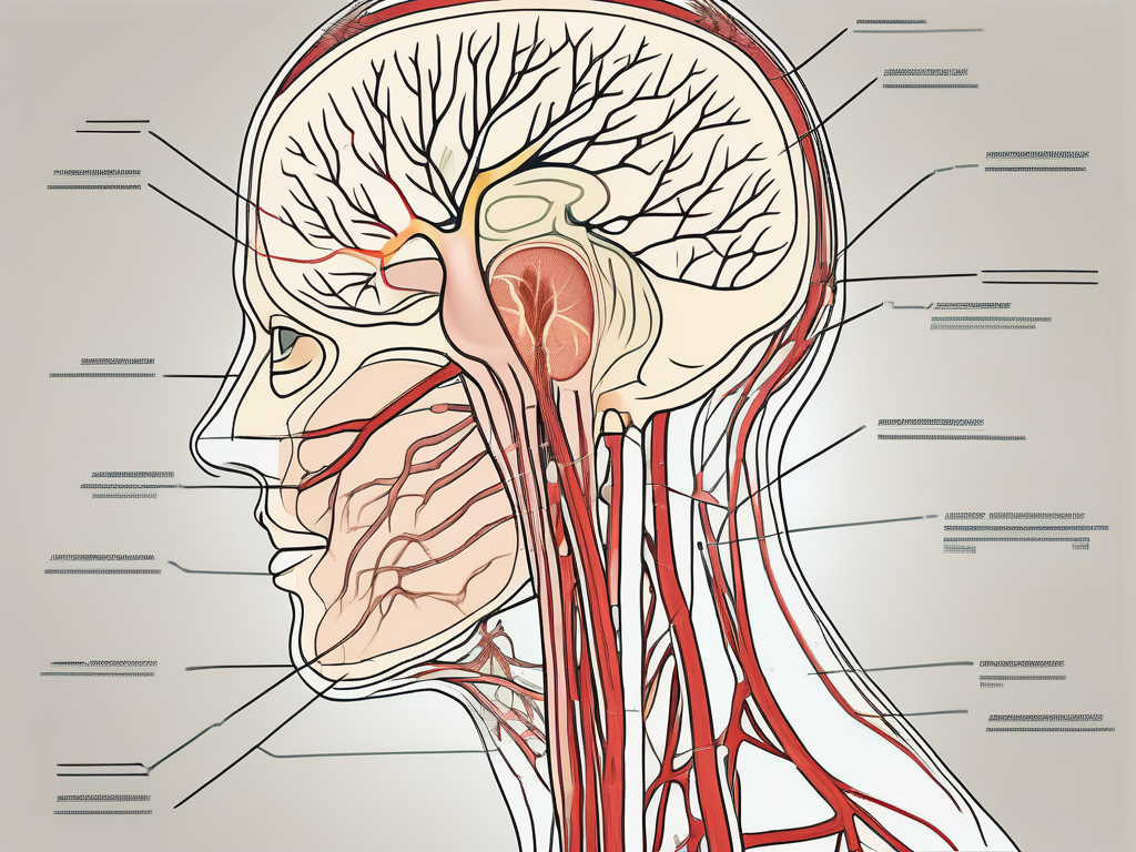
The abducens nerve arises from the pons, a region in the brainstem that serves as a vital connection between the brain and the spinal cord. It extends towards the muscles controlling lateral eye movement on each side of the head.
As the abducens nerve follows its pathway, it navigates through a complex and fascinating anatomical arrangement. It crosses the skull through the cavernous sinus, a cavity located behind the eyes. This intricate journey allows the abducens nerve to establish precise control over eye movements, ensuring coordinated and accurate shifts in our visual field.
Function of the Abducens Nerve
The primary function of the abducens nerve is to facilitate the abduction of the eye. Abduction refers to the lateral movement away from the center, enabling us to explore our surroundings and focus on objects located to the side.
By activating the muscle it innervates, the abducens nerve initiates the movement required for eye abduction. This coordinated effort allows us to effortlessly scan our environment, capturing important details and expanding our visual perception.
Furthermore, the abducens nerve works in harmony with other cranial nerves and structures in the brain responsible for eye movement. This collaboration ensures proper alignment and smooth motions, preventing any disruptions or misalignment that may hinder our visual experience.
Understanding the abducens nerve and its intricate role in eye movement provides us with a glimpse into the remarkable complexity of the human body. The precise control and coordination required for even the simplest of eye movements highlight the intricate nature of our anatomy and the wonders of our nervous system.
The Muscle Innervated by the Abducens Nerve
Identifying the Muscle
The abducens nerve innervates the lateral rectus muscle, one of the extraocular muscles situated in the eye socket. This muscle, as its name implies, is responsible for moving the eye away from the center of the face. Its contraction enables the eyeball to gaze outward, allowing for a wider field of view.
The lateral rectus muscle works in opposition to the medial rectus muscle, which is innervated by the oculomotor nerve and performs inward eye movements.
Role and Function of the Muscle
In conjunction with the abducens nerve, the lateral rectus muscle ensures precise coordination of eye movements, contributing to binocular vision and depth perception. By working synergistically with other ocular muscles, the lateral rectus muscle enables the eyes to move synchronously, facilitating various visual tasks.
Furthermore, the lateral rectus muscle plays a critical role in maintaining eye alignment, preventing conditions such as strabismus, where the eyes do not focus in the same direction. Proper functioning of this muscle is essential for normal eye movement and optimal visual function.
When the abducens nerve sends signals to the lateral rectus muscle, it triggers a series of events that result in the contraction of the muscle fibers. These muscle fibers are composed of specialized cells called myocytes, which have the unique ability to generate force and produce movement.
Within the lateral rectus muscle, there are numerous nerve endings that receive signals from the abducens nerve. These nerve endings transmit electrical impulses, which stimulate the muscle fibers to contract. The contraction of the lateral rectus muscle causes the eyeball to rotate outward, allowing the individual to look to the side.
It is important to note that the lateral rectus muscle does not work in isolation. It is part of a complex network of muscles and nerves that work together to control eye movements. The abducens nerve, along with other cranial nerves, plays a crucial role in coordinating the actions of these muscles, ensuring smooth and accurate eye movements.
Additionally, the lateral rectus muscle is surrounded by connective tissue, which provides support and protection. This tissue helps to maintain the structural integrity of the muscle, allowing it to function optimally. Without the presence of this connective tissue, the lateral rectus muscle would be more susceptible to injury and damage.
In summary, the abducens nerve innervates the lateral rectus muscle, which is responsible for moving the eye outward. This muscle plays a vital role in coordinating eye movements, maintaining eye alignment, and facilitating various visual tasks. Understanding the intricate details of the anatomy and function of the lateral rectus muscle provides valuable insights into the complex mechanisms that govern eye movement and visual perception.
The Connection between the Abducens Nerve and the Muscle
How the Abducens Nerve Controls the Muscle
The abducens nerve, also known as cranial nerve VI, plays a crucial role in controlling the movement of the eye. Specifically, it communicates with the lateral rectus muscle through motor neurons, ensuring efficient transmission of signals from the brain to the muscle fibers. This intricate connection allows for precise control of eye movement.
When the abducens nerve is stimulated, it sends electrical impulses to the lateral rectus muscle, prompting its contraction. This contraction, in turn, causes the eye to move away from the midline, towards the outer side of the face. This outward movement is known as abduction, hence the name of the nerve.
It is fascinating to consider the complexity of this process. The abducens nerve acts as a messenger, relaying information from the brain to the muscle, ultimately resulting in the coordinated movement of the eye. This precise coordination enables the eyes to work in harmony, facilitating accurate tracking of moving objects and enhancing our visual experiences.
The Impact of the Abducens Nerve on Muscle Movement
The abducens nerve’s influence on the lateral rectus muscle is of paramount importance. Any disruption in its function can lead to altered eye movement patterns and associated visual difficulties. Disorders affecting the abducens nerve can result in limited or impaired lateral eye movement, leading to potential vision disturbances.
One such disorder is called abducens nerve palsy, which occurs when the abducens nerve is damaged or compressed. This condition can cause weakness or paralysis of the lateral rectus muscle, making it difficult for the affected eye to move outward. As a result, individuals with abducens nerve palsy may experience double vision, especially when looking towards the affected side.
It is imperative to monitor any changes in eye movement and consult with a healthcare professional if persistent or concerning issues arise. Proper diagnosis and treatment are essential to address abducens nerve-related disorders effectively. In some cases, surgical intervention may be necessary to restore normal function and alleviate symptoms.
Understanding the intricate connection between the abducens nerve and the lateral rectus muscle highlights the incredible complexity of the human body. The delicate interplay between nerves and muscles allows us to perform precise movements and perceive the world around us. By appreciating the significance of this connection, we can better understand and appreciate the wonders of our visual system.
Disorders Related to the Abducens Nerve
The abducens nerve, also known as the sixth cranial nerve, plays a crucial role in eye movement. When this nerve is affected by various disorders, it can lead to a range of symptoms that can significantly impact a person’s vision and overall quality of life.
Symptoms of Abducens Nerve Disorders
Abducens nerve disorders can manifest in various symptoms, each with its own unique impact on visual function. One of the most common symptoms is double vision, also known as diplopia. This occurs when the eyes are unable to align properly, resulting in the perception of two images instead of one.
In addition to double vision, individuals with abducens nerve disorders may experience difficulty moving their eyes laterally. This can make it challenging to focus on objects located to the side, affecting tasks such as reading, driving, or even engaging in conversations.
Eye misalignment, another common symptom, can cause one eye to deviate inward or outward, resulting in a condition known as strabismus. This not only affects the appearance of the eyes but can also lead to further visual disturbances and a loss of depth perception.
Eye fatigue is another symptom associated with abducens nerve disorders. The constant strain on the eyes due to misalignment or difficulty in eye movement can lead to feelings of tiredness, heaviness, and discomfort. This can significantly impact daily activities that require visual concentration, such as working on a computer or reading for extended periods.
Headaches associated with eye movement are also commonly reported by individuals with abducens nerve disorders. These headaches can range from mild to severe and may be localized around the eyes or spread to other areas of the head. The pain is often exacerbated by eye movement, making it difficult to perform even simple tasks.
If you experience any of these symptoms, it is important to seek medical attention promptly. Early diagnosis and intervention can help prevent further complications and improve the overall prognosis.
Diagnosis and Treatment of Abducens Nerve Disorders
Consultation with a healthcare professional, such as a neurologist or ophthalmologist, is crucial for the diagnosis and management of abducens nerve disorders. These specialists have the expertise to evaluate your symptoms, perform a comprehensive examination, and order additional tests to identify the underlying cause of your condition.
Neuroimaging studies, such as magnetic resonance imaging (MRI) or computed tomography (CT) scans, may be recommended to assess the structure and function of the brain and surrounding structures. These imaging techniques can provide valuable insights into potential abnormalities that may be affecting the abducens nerve.
Once a diagnosis is made, the appropriate course of treatment can be determined. Treatment options for abducens nerve disorders depend on the specific diagnosis and severity of the condition. In some cases, therapeutic exercises may be prescribed to strengthen the eye muscles and improve coordination. Eye patching or the use of prism glasses can also be beneficial in certain cases to help correct misalignment and alleviate symptoms.
Medication may be prescribed to manage underlying conditions contributing to the abducens nerve disorder, such as inflammation or nerve damage. In severe instances where conservative measures fail to provide relief, surgical intervention may be considered. Surgical procedures aim to correct structural abnormalities or address any issues affecting the abducens nerve directly.
It is essential to follow your healthcare professional’s guidance and recommendations to ensure the best possible outcomes. Regular follow-up appointments and ongoing monitoring of your condition may be necessary to track progress and make any necessary adjustments to the treatment plan.
Remember, early intervention and proper management of abducens nerve disorders can significantly improve visual function and enhance overall quality of life.
Maintaining the Health of the Abducens Nerve
The abducens nerve plays a crucial role in eye movement, specifically in the lateral rectus muscle. Keeping this nerve healthy is essential for maintaining proper ocular function. While some abducens nerve disorders may be unavoidable, certain preventive measures can help maintain its health and functionality.
Preventive Measures for Abducens Nerve Health
Adopting a healthy lifestyle is one of the key factors in promoting the health of the abducens nerve. This includes maintaining a balanced diet that is rich in essential nutrients. Nutrients like vitamin A, omega-3 fatty acids, and antioxidants can support optimal nerve function and overall eye health.
Regular exercise is also beneficial for the abducens nerve. Engaging in physical activities that promote blood circulation can help nourish the nerve and keep it in good condition. Additionally, exercise can help reduce the risk of developing conditions that could affect the abducens nerve and overall ocular function.
Adequate rest is another crucial aspect of maintaining the health of the abducens nerve. Getting enough sleep allows the body to repair and regenerate, including the nerves. It is recommended to aim for 7-9 hours of quality sleep each night to support optimal nerve function.
Protecting the eyes from trauma is essential in preventing damage to the abducens nerve. Wearing protective eyewear during activities that pose a risk of eye injury, such as sports or certain occupations, can significantly reduce the chances of nerve-related complications.
Furthermore, avoiding excessive strain on the eyes is crucial for maintaining the health of the abducens nerve. Prolonged periods of screen time, whether it be from computers, smartphones, or other digital devices, can strain the eyes and potentially impact nerve function. Taking regular breaks and practicing proper eye care, such as the 20-20-20 rule (looking away from the screen every 20 minutes and focusing on something 20 feet away for 20 seconds), can help alleviate strain on the abducens nerve.
Rehabilitation and Exercises for the Abducens Nerve and Muscle
For individuals recovering from abducens nerve-related disorders, rehabilitation programs supervised by healthcare professionals can play a crucial role in the recovery process. These programs may incorporate specific exercises targeting the affected muscle and related brain-eye coordination.
Exercises that focus on eye movements, such as controlled eye tracking and gaze stabilization exercises, can help restore proper function and improve visual outcomes. These exercises aim to strengthen the abducens nerve and promote coordination between the brain and the affected muscle.
It is essential to consult with a healthcare professional experienced in vision-related rehabilitation, such as a physical therapist or occupational therapist. They can assess your specific condition and develop an appropriate exercise plan tailored to your needs. These professionals can guide you through the exercises, ensuring proper technique and progression for optimal recovery.
As with any medical concern, it is essential to seek professional medical advice and guidance. The information presented in this article aims to provide a general understanding of the abducens nerve and its relation to the muscle it innervates, as well as disorders and preventive measures. However, every individual’s situation is unique, and your healthcare provider can provide personalized recommendations and address any concerns you may have regarding your specific condition.
Leave a Reply