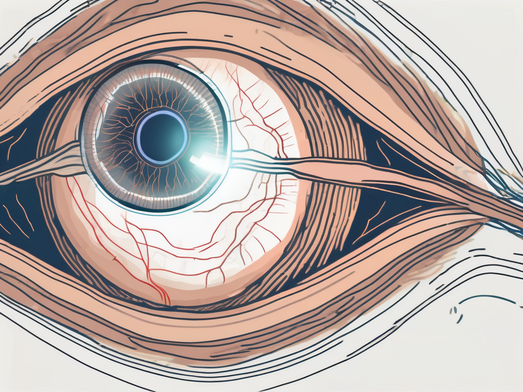
The abducens nerve, also known as the sixth cranial nerve, plays a crucial role in the movement of the eye. It controls the action of the lateral rectus muscle, which is responsible for the sideways movement of the eyeball. Understanding the anatomy and functioning of both the abducens nerve and the lateral rectus muscle is essential in comprehending the complex mechanisms involved in eye movement.
Understanding the Anatomy of the Eye
Eyes are remarkable organs that allow us to perceive the world around us. The intricate structure of the eye consists of various components working together to achieve clear and focused vision. Among these components are the muscles that control the movement of the eyeball.
The muscles responsible for moving the eye are known as extraocular muscles. These muscles work in perfect harmony to allow for precise movements in various directions. There are six extraocular muscles surrounding each eyeball, each with its own specific function. The superior rectus muscle, for example, is responsible for moving the eye upward, while the inferior rectus muscle moves it downward. The lateral rectus muscle allows for side-to-side movement, and the medial rectus muscle brings the eye inward towards the nose. Additionally, the superior oblique muscle helps to rotate the eye downward and inward, while the inferior oblique muscle rotates it upward and outward.
The Role of Muscles in Eye Movement
Eye movement is not solely reliant on the muscles; however, they are crucial for directing the eyes towards a specific object or point of interest. These muscles work in perfect coordination to ensure that our gaze is accurately directed. For example, when we read a book, our eyes move smoothly across the page, thanks to the precise control of these muscles. Without them, our eyes would be unable to track moving objects or focus on different points in our visual field.
Interestingly, the extraocular muscles are among the fastest and most precise muscles in the human body. They can make rapid movements, allowing us to quickly shift our gaze from one object to another. This incredible speed and accuracy are essential for activities such as playing sports, driving, or even just following a moving object.
The Importance of Nerves in Eye Functioning
Nerves play a vital role in transmitting signals between the brain and the muscles, allowing for coordinated eye movements. These signals travel along a complex network of nerves, starting from the visual cortex in the brain and reaching the extraocular muscles. This intricate system ensures that the brain can control the eye movements with incredible precision.
Without the proper functioning of these nerves, eye movements can be impaired, leading to various eye-related conditions. For example, damage to the nerves that control the extraocular muscles can result in strabismus, a condition where the eyes are misaligned and do not point in the same direction. This can cause double vision and difficulties with depth perception. Additionally, conditions like nystagmus, where the eyes make involuntary and repetitive movements, can also be attributed to nerve dysfunction.
It is fascinating to think about how the muscles and nerves work together seamlessly to allow us to see the world around us. The complexity of the eye’s anatomy and its intricate connections with the brain highlight the incredible design of our visual system. Understanding the role of muscles and nerves in eye functioning not only deepens our appreciation for the wonders of the human body but also helps us comprehend the various eye conditions that can arise when these components are not functioning optimally.
The Abducens Nerve: An Overview
The abducens nerve, also referred to as cranial nerve VI, originates in the pons of the brainstem. It is responsible for controlling the lateral rectus muscle, which moves the eye away from the midline of the face.
Origin and Pathway of the Abducens Nerve
The abducens nerve emerges from the brainstem and travels along a complex pathway towards the eye muscles. This nerve takes a unique route that distinguishes it from other cranial nerves, passing through crucial structures and areas within the skull.
As the abducens nerve exits the brainstem, it passes through the cavernous sinus, a large venous channel located on each side of the sella turcica, a bony saddle-like structure in the skull. This sinus is a complex network of veins that serves as a conduit for multiple cranial nerves and blood vessels.
Continuing its journey, the abducens nerve then enters the superior orbital fissure, a narrow opening located in the sphenoid bone. This fissure serves as a pathway for various structures, including the abducens nerve, to reach the orbit of the eye.
Upon entering the orbit, the abducens nerve finally reaches its destination, the lateral rectus muscle. This muscle is one of the six extraocular muscles responsible for eye movement. The abducens nerve innervates the lateral rectus muscle, allowing it to contract and move the eyeball laterally.
Functions of the Abducens Nerve
The primary function of the abducens nerve is to innervate the lateral rectus muscle, enabling it to contract and move the eyeball laterally. This allows for horizontal movements of the eyes, which are essential for gaze shifts and tracking moving objects.
When the abducens nerve is functioning properly, both eyes work together to create a unified visual perception. However, if there is a dysfunction or damage to the abducens nerve, it can lead to a condition called abducens nerve palsy. This condition results in the inability to move the affected eye laterally, causing double vision and difficulty with eye coordination.
In addition to its role in eye movement, the abducens nerve also plays a crucial role in maintaining eye alignment. It works in conjunction with other cranial nerves and muscles to ensure that both eyes are properly aligned, allowing for binocular vision and depth perception.
Furthermore, the abducens nerve is involved in the vestibulo-ocular reflex, which helps stabilize the eyes during head movements. This reflex allows the eyes to move in the opposite direction to compensate for head movements, ensuring that the visual field remains stable.
In conclusion, the abducens nerve is a vital component of the intricate network responsible for eye movement and coordination. Its unique pathway and functions highlight its importance in maintaining proper eye alignment and visual perception.
The Lateral Rectus Muscle: The Muscle Supplied by the Abducens Nerve
The lateral rectus muscle, as the name suggests, is one of the six extraocular muscles that operate the eyeball’s movement. It is unique as it is solely controlled by the abducens nerve, which enables it to perform its specific function.
Located on the outer side of the eyeball, the lateral rectus muscle plays a crucial role in the intricate system of eye movements. Its attachment to the eyeball allows it to exert a pulling force, directing the eye towards the temple. This action results in the eye rotating outwardly, away from the midline of the body.
Working in harmony with the other extraocular muscles, the lateral rectus muscle ensures smooth and coordinated eye movements. These muscles, including the superior rectus, inferior rectus, medial rectus, superior oblique, and inferior oblique, work together to enable the eye to move in various directions.
Anatomy of the Lateral Rectus Muscle
The lateral rectus muscle is a slender and flat muscle that originates from a small bony prominence called the annulus of Zinn. From there, it extends horizontally across the eye socket and attaches to the outer side of the eyeball. This specific attachment allows the lateral rectus muscle to exert its pulling force effectively, enabling the eye to rotate outwardly.
Like other muscles in the body, the lateral rectus muscle is composed of bundles of muscle fibers. These fibers are innervated by the abducens nerve, also known as cranial nerve VI. This nerve originates in the brainstem and travels through the skull to reach the lateral rectus muscle, providing it with the necessary signals for contraction and relaxation.
Role of the Lateral Rectus Muscle in Eye Movement
The lateral rectus muscle’s primary function is to abduct the eye, meaning it moves the eye away from the nose. This muscle works in opposition to the medial rectus muscle, which brings the eye back towards the midline. The coordinated actions of these muscles allow for horizontal eye movements.
Imagine looking at a distant object to your right. In this scenario, the lateral rectus muscle contracts, pulling the eyeball towards the temple, allowing you to focus on the object. On the other hand, when you shift your gaze towards the midline, the medial rectus muscle takes over, bringing the eye back to its original position.
These intricate movements of the eye are essential for various activities, such as reading, driving, and following objects in our visual field. Without the precise functioning of the lateral rectus muscle, our ability to explore the world visually would be greatly impaired.
The Relationship between the Abducens Nerve and the Lateral Rectus Muscle
The abducens nerve and the lateral rectus muscle share an intricate and mutually dependent relationship. The nerve provides the necessary signals for the muscle to contract, resulting in the movement of the eyeball.
The abducens nerve, also known as cranial nerve VI, is responsible for the innervation of the lateral rectus muscle. This muscle is one of the six extraocular muscles that control the movement of the eye. Its primary function is to abduct the eye, meaning it moves the eyeball away from the midline of the face.
The lateral rectus muscle is located on the outer side of each eye, opposite the medial rectus muscle. These two muscles work in opposition to each other, allowing for coordinated eye movements. When the abducens nerve sends signals to the lateral rectus muscle, it contracts, pulling the eyeball outward.
How the Abducens Nerve Controls the Lateral Rectus Muscle
The abducens nerve communicates with the lateral rectus muscle through a specialized neuromuscular junction. This junction is the point where the nerve and muscle meet, allowing for the transmission of signals. When the nerve sends a signal, the neurotransmitter acetylcholine is released, initiating muscle contraction. This coordinated process ensures precise and controlled eye movements.
It is important to note that the abducens nerve not only controls the lateral rectus muscle but also receives feedback from it. This feedback loop helps maintain the correct positioning and alignment of the eyes, allowing for binocular vision and depth perception.
In addition to the abducens nerve, other cranial nerves, such as the oculomotor nerve (cranial nerve III), play a role in eye movement. These nerves work together to ensure smooth and coordinated eye movements in various directions.
Consequences of Damage to the Abducens Nerve on Eye Movement
Dysfunction or damage to the abducens nerve can lead to various conditions, such as abducens nerve palsy. This condition can result in an impaired ability to move the affected eye laterally, leading to difficulties in maintaining binocular vision and accurate eye coordination.
Abducens nerve palsy can be caused by various factors, including trauma, infections, tumors, or neurological disorders. Depending on the severity of the damage, individuals with abducens nerve palsy may experience double vision, eye misalignment, or limited eye movement.
Treatment for abducens nerve palsy depends on the underlying cause and may include medications, eye exercises, or in some cases, surgery. Rehabilitation and visual therapy can also be beneficial in improving eye coordination and restoring normal eye movements.
In conclusion, the relationship between the abducens nerve and the lateral rectus muscle is crucial for the precise control of eye movements. The coordinated signals between the nerve and muscle allow for smooth and accurate lateral eye movements, contributing to our ability to perceive the world around us.
Medical Conditions Related to the Abducens Nerve and Lateral Rectus Muscle
The abducens nerve and lateral rectus muscle play a crucial role in the functioning and movement of the eye. Understanding the relationship between these two components is essential in diagnosing and treating certain eye conditions that may arise from dysfunction in this complex system.
The abducens nerve, also known as the sixth cranial nerve, is responsible for innervating the lateral rectus muscle. This muscle is one of the six extraocular muscles that control the movement of the eye. Its primary function is to abduct or move the eye laterally, allowing for horizontal eye movements.
When there is dysfunction in the abducens nerve or the lateral rectus muscle, it can lead to various medical conditions. One such condition is abducens nerve palsy, which is characterized by a weakened or paralyzed lateral rectus muscle. This results in limited or absent abduction of the affected eye.
Symptoms and Diagnosis of Abducens Nerve Palsy
Abducens nerve palsy can cause a range of symptoms that can significantly impact a person’s vision and eye movements. Individuals with this condition may experience double vision, also known as diplopia, where they see two images instead of one. This occurs because the affected eye is unable to align properly with the other eye, leading to misalignment or strabismus.
In addition to double vision, individuals with abducens nerve palsy may have difficulty with certain eye movements. They may find it challenging to move their affected eye laterally, resulting in limited side-to-side eye movements. This can affect their ability to track moving objects or scan their surroundings effectively.
Diagnosing abducens nerve palsy typically involves a comprehensive eye examination and evaluation. An ophthalmologist or neurologist will assess the patient’s eye movements, visual acuity, and perform specialized tests to determine the underlying cause of the condition. Imaging studies, such as magnetic resonance imaging (MRI), may also be used to identify any structural abnormalities or lesions affecting the abducens nerve or lateral rectus muscle.
Treatment Options for Abducens Nerve and Lateral Rectus Muscle Disorders
The treatment options for abducens nerve and lateral rectus muscle disorders depend on the underlying cause and severity of the condition. It is crucial for individuals experiencing symptoms to consult with a qualified ophthalmologist or neurologist who specializes in eye conditions to determine the most appropriate treatment plan.
In some cases, abducens nerve palsy may resolve on its own without intervention. However, if the condition persists or significantly affects a person’s vision and daily activities, treatment may be necessary. Treatment options can include medication, therapies, or, in severe cases, surgery.
Medication may be prescribed to manage any underlying medical conditions contributing to the dysfunction of the abducens nerve or lateral rectus muscle. For example, if the palsy is caused by nerve inflammation, anti-inflammatory medications may be prescribed to reduce swelling and promote healing.
Therapies such as vision therapy or eye exercises can also be beneficial in improving eye muscle coordination and strengthening the affected muscles. These therapies aim to enhance the brain-eye muscle connection and restore proper eye movements.
In rare cases where conservative treatments are ineffective or the condition is severe, surgery may be considered. Surgical intervention may involve procedures to correct muscle alignment, strengthen weak eye muscles, or address any structural abnormalities affecting the abducens nerve.
Understanding the intricate interplay between the abducens nerve and the lateral rectus muscle is essential in comprehending the functioning and movement of the eye. While this article provides a general overview, it is important to consult with a medical professional for any concerns or questions relating to specific eye conditions or symptoms. Only by seeking appropriate medical advice can an accurate diagnosis and treatment plan be established for optimal eye health and functioning.

Leave a Reply