The abducens nerve, also known as the sixth cranial nerve, is a vital component of the human nervous system. It plays a crucial role in controlling eye movement by innervating specific regions of the ocular muscles. Understanding the anatomy and function of the abducens nerve is essential to appreciate its significance and the potential disorders associated with it.
Understanding the Abducens Nerve
The abducens nerve, also known as cranial nerve VI, plays a crucial role in eye movement and coordination. It is responsible for the lateral movement of the eyes, allowing controlled abduction or outward rotation. Let’s delve deeper into the anatomy and function of this fascinating nerve.
Anatomy of the Abducens Nerve
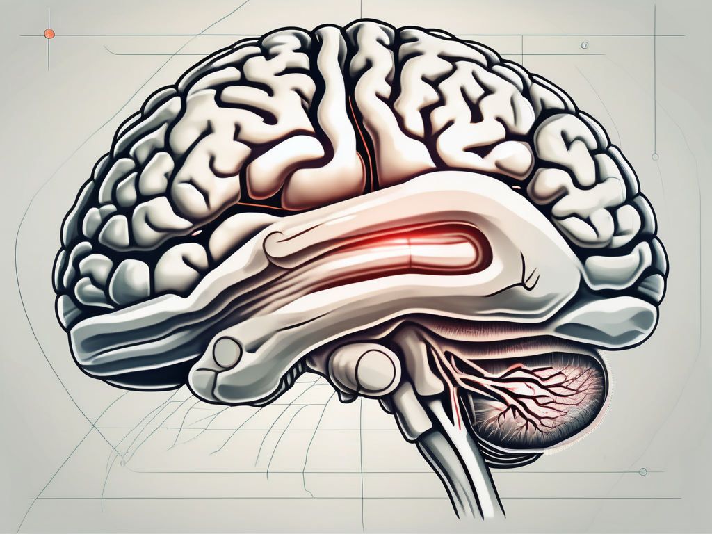
The abducens nerve arises from the pons, a part of the brainstem located in the posterior fossa of the skull. It emerges from the brainstem along with other cranial nerves, forming the cranial nerve nuclei. From its origin, the abducens nerve travels through the cavernous sinus, a cavity located on each side of the sella turcica, a bony structure in the skull.
As the abducens nerve courses through the cavernous sinus, it navigates its way to reach the lateral rectus muscle in each eye. The lateral rectus muscle is one of the six extraocular muscles responsible for eye movement. It is located on the outer side of each eye and is primarily responsible for abducting or moving the eye laterally.
The motor fibers originating from the abducens nucleus, located in the pons, branch out to innervate the lateral rectus muscle. These motor fibers transmit electrical signals from the brain to the muscle, allowing precise control over the abduction or outward rotation of the eyes.
Function of the Abducens Nerve
The abducens nerve plays a vital role in ensuring normal eye movement and coordination. By providing input to the lateral rectus muscle, it enables horizontal gaze and contributes to binocular vision. Binocular vision refers to the ability of both eyes to work together, providing a three-dimensional perception of the surrounding environment.
Working in harmony with other cranial nerves, such as the oculomotor nerve (cranial nerve III) and the trochlear nerve (cranial nerve IV), the abducens nerve helps in maintaining the precise alignment of the eyes. This alignment ensures that both eyes focus on the same object, allowing for simultaneous visual input and reduced visual disturbances.
Any disruption or damage to the abducens nerve can lead to a condition called abducens nerve palsy. Abducens nerve palsy results in the inability to move the affected eye laterally, causing double vision (diplopia) and difficulties in focusing on objects to the side. This condition can be caused by various factors, including trauma, infections, tumors, or neurological disorders.
In conclusion, the abducens nerve is a vital component of the intricate system that governs eye movement and coordination. Its role in controlling the lateral movement of the eyes and contributing to binocular vision is crucial for our daily visual experiences.
The Region Innervated by the Abducens Nerve
The abducens nerve, also known as the sixth cranial nerve, is responsible for innervating the lateral rectus muscle of the eye. This muscle is crucial for horizontal eye movement, allowing the eyes to move outward. The abducens nerve plays a vital role in the voluntary control of eye movement, facilitating smooth tracking of moving objects and exploration of the visual field.
When the abducens nerve is functioning properly, it ensures coordinated eye movements, enhancing visual perception and spatial awareness. This coordination is particularly important for activities such as reading, driving, and playing sports, where precise eye movements are necessary for optimal performance.
Role in Eye Movement
The abducens nerve’s primary role is to facilitate the outward movement of the eyes. This movement is known as abduction, hence the name of the nerve. By contracting the lateral rectus muscle, the abducens nerve allows the eyes to move away from the midline, enabling binocular vision and expanding the visual field.
Smooth eye movements are essential for maintaining focus on a moving object or tracking multiple objects simultaneously. The abducens nerve’s involvement in horizontal gaze ensures that both eyes work together harmoniously, allowing for accurate eye movements and enhancing visual tracking abilities.
In addition to its role in voluntary eye movements, the abducens nerve also contributes to the vestibulo-ocular reflex. This reflex helps stabilize the eyes during head movements, ensuring that the visual field remains steady even when the head is in motion.
Connection to the Brainstem
The abducens nerve originates from the abducens nucleus, which is located within the pons of the brainstem. The brainstem is a vital part of the central nervous system, connecting the brain to the spinal cord and facilitating communication between different regions of the nervous system.
The abducens nucleus contains the cell bodies of the abducens nerve fibers. These fibers exit the brainstem and travel through the cavernous sinus, a cavity located behind the eyes. From there, the nerve fibers reach the lateral rectus muscle, innervating it and allowing for its contraction.
This intricate connection between the abducens nucleus and the lateral rectus muscle ensures precise control over eye movements. Dysfunction or damage to the abducens nucleus can have profound effects on eye movement and coordination, leading to various disorders such as strabismus (crossed eyes) or diplopia (double vision).
Understanding the region innervated by the abducens nerve and its connection to the brainstem is crucial for diagnosing and treating eye movement disorders. By studying the intricate anatomy and physiology of this nerve, researchers and healthcare professionals can develop targeted interventions to improve eye movement control and enhance visual function.
Disorders Related to the Abducens Nerve
The abducens nerve plays a crucial role in the movement of the eye. When this nerve is damaged or dysfunctional, it can lead to a condition known as abducens nerve palsy. This condition is characterized by weakness or paralysis of the lateral rectus muscle, which is responsible for moving the eye towards the outer periphery.
Symptoms of Abducens Nerve Palsy
Abducens nerve palsy can cause a range of symptoms that can significantly impact a person’s vision and daily life. One of the most common symptoms is lateral eye misalignment, where the affected eye deviates inward, causing double vision. This can make it challenging to focus on objects and can lead to difficulties in depth perception.
In addition to eye misalignment and double vision, individuals with abducens nerve palsy may also experience difficulty in moving the affected eye towards the outer periphery. This limitation in eye movement can make it challenging to track objects or follow a moving target.
If you are experiencing any of these symptoms, it is crucial to seek medical attention. While some cases of abducens nerve palsy may resolve on their own, persistent or worsening symptoms may indicate an underlying issue that requires treatment.
Treatment and Recovery Options
The treatment and recovery options for abducens nerve palsy depend on various factors, including the underlying cause and the severity of the condition. In mild cases, conservative management techniques may be sufficient to alleviate symptoms. These techniques can include patching, where the unaffected eye is covered to encourage the affected eye to strengthen and regain its functionality. Another option is the use of prisms, which can help correct double vision by redirecting light rays to align properly.
However, in more severe cases of abducens nerve palsy, surgical intervention or targeted therapies may be necessary. Surgery can involve procedures such as muscle repositioning or tightening, which aim to restore proper eye alignment and improve eye movement. Targeted therapies, such as botulinum toxin injections, can also be used to temporarily weaken specific eye muscles, allowing for better alignment and reducing double vision.
It is important to consult with a healthcare professional to determine the most suitable treatment approach for your specific case of abducens nerve palsy. They will consider factors such as the underlying cause, your overall health, and your individual needs to develop a personalized treatment plan.
While the road to recovery may vary for each individual, with proper treatment and management, many people with abducens nerve palsy can experience significant improvement in their symptoms. Rehabilitation exercises, such as eye muscle strengthening and coordination exercises, may also be recommended to aid in the recovery process.
Remember, early intervention and regular follow-up with your healthcare provider are essential in managing abducens nerve palsy effectively. By seeking timely medical attention and adhering to the recommended treatment plan, you can increase your chances of a successful recovery and minimize the impact of this condition on your daily life.
The Abducens Nerve in the Medical Field
The abducens nerve plays a crucial role in the field of medicine, particularly in the assessment and treatment of various conditions. This nerve, also known as the sixth cranial nerve, is responsible for the movement of the lateral rectus muscle, which controls the outward movement of the eye. Dysfunction or damage to the abducens nerve can lead to a range of symptoms, including double vision and difficulty moving the affected eye.
Diagnostic Procedures Involving the Abducens Nerve
Medical professionals employ a variety of diagnostic procedures to evaluate the function and integrity of the abducens nerve. These procedures are essential in determining the underlying cause of any abnormalities and formulating appropriate treatment plans.
One of the primary diagnostic tools used is a detailed neurological examination. This examination involves a comprehensive assessment of the patient’s eye movements, including the ability to move the eyes laterally. By carefully observing the patient’s eye movements, medical professionals can identify any abnormalities or restrictions that may be indicative of abducens nerve dysfunction.
In addition to the neurological examination, imaging studies such as magnetic resonance imaging (MRI) or computed tomography (CT scan) may be performed. These imaging techniques provide detailed images of the brain and cranial structures, allowing medical professionals to visualize the abducens nerve and identify any potential lesions or compressions that may be affecting its function.
Specialized tests, such as electrodiagnostic studies, may also be utilized to assess the function of the abducens nerve. These tests involve the measurement of electrical signals generated by the nerve, providing valuable information about its integrity and responsiveness.
By combining the findings from these diagnostic procedures, medical professionals can accurately diagnose abducens nerve disorders and develop targeted treatment plans. This comprehensive approach ensures that patients receive the most appropriate and effective care for their specific condition.
Surgical Considerations and the Abducens Nerve
During certain surgical procedures, particularly those involving the brainstem or cranial base, surgeons must exercise extreme caution to protect the delicate abducens nerve. The close proximity of this nerve to critical structures necessitates meticulous planning and precise execution to avoid inadvertent damage.
Prior to any surgical intervention, a thorough preoperative assessment is conducted to evaluate the anatomical relationship between the abducens nerve and surrounding structures. This assessment often involves collaboration between neurosurgeons, otolaryngologists, and ophthalmologists to ensure a comprehensive understanding of the surgical site.
During surgery, various techniques may be employed to minimize the risk of abducens nerve injury. Surgeons may use intraoperative monitoring, which involves the real-time assessment of nerve function, to guide their surgical maneuvers and avoid any inadvertent damage to the nerve.
Additionally, the use of advanced imaging techniques, such as intraoperative MRI or CT scans, may provide real-time visualization of the abducens nerve during surgery. This allows surgeons to precisely identify and navigate around the nerve, further reducing the risk of injury.
Overall, the careful consideration and protection of the abducens nerve during surgical procedures are paramount to ensure optimal patient outcomes. The collaboration between different medical specialties and the utilization of advanced technologies contribute to the success and safety of these complex interventions.
The Abducens Nerve in Research
The abducens nerve, also known as the sixth cranial nerve, is a critical component of the human visual system. It is responsible for controlling the movement of the lateral rectus muscle, which allows the eye to move laterally or outward. Ongoing research has focused on unraveling the intricate mechanisms and connections involved in abducens nerve function, leading to significant advancements in our understanding of this vital cranial nerve.
Recent Discoveries about the Abducens Nerve
Recent studies have shed light on various aspects of the abducens nerve, revealing fascinating insights into its development, maintenance, and function. One area of research has focused on the role of genes in the formation and regulation of the abducens nerve. Scientists have identified specific genes that play a crucial role in guiding the growth of the nerve fibers and ensuring their proper connectivity with the brainstem.
Furthermore, researchers have explored the molecular pathways involved in the development and maintenance of the abducens nerve. By studying the intricate signaling mechanisms within the nerve cells, scientists have gained a deeper understanding of how these cells communicate and coordinate their activities to ensure precise eye movement control.
In addition to genetic and molecular studies, advancements in imaging techniques have revolutionized our ability to visualize and study the abducens nerve’s anatomical characteristics. High-resolution imaging methods, such as magnetic resonance imaging (MRI) and diffusion tensor imaging (DTI), have allowed researchers to map the precise trajectory of the abducens nerve fibers within the brainstem. This detailed anatomical information has provided valuable insights into the nerve’s connectivity with other brain regions and its relationship with adjacent cranial nerves.
Future Directions for Abducens Nerve Research
The future of abducens nerve research holds great promise for further unraveling the complexities of this crucial cranial nerve. One exciting avenue of investigation is exploring the intricate relationship between the abducens nerve and other cranial nerves involved in eye movement control. By studying the interactions and coordination between these nerves, researchers hope to gain a comprehensive understanding of the neural circuitry underlying eye movement.
Moreover, ongoing investigations into the molecular, cellular, and genetic aspects of the abducens nerve may lead to the discovery of novel therapeutic targets and interventions for related disorders. Understanding the underlying mechanisms of abducens nerve dysfunction could pave the way for the development of targeted treatments that could restore normal eye movement and coordination in individuals with abducens nerve disorders.
Collaboration between clinicians and researchers is crucial for translating scientific advancements into improved patient care and outcomes. By working together, medical professionals can provide valuable clinical insights and feedback to researchers, guiding their investigations towards addressing the most pressing clinical challenges associated with abducens nerve dysfunction.
In conclusion, the abducens nerve is a fascinating area of research that continues to reveal new insights into its anatomy, function, and associated disorders. The ongoing efforts of scientists and clinicians in studying this vital cranial nerve hold great promise for improving our understanding of eye health and developing innovative treatments for individuals with abducens nerve-related conditions. It is essential to consult with a qualified healthcare provider for personalized advice and treatment options should any concerns arise regarding the function or integrity of the abducens nerve.
Leave a Reply