The abducens nerve, also known as the sixth cranial nerve or cranial nerve VI, plays a critical role in the movement of the eye. Understanding the anatomy, function, and synaptic process of this nerve is essential in comprehending the complex mechanisms behind ocular control and the various disorders related to it.
Understanding the Abducens Nerve
The abducens nerve is a crucial component of the ocular motor system, responsible for controlling the movement of the eye. This nerve plays a vital role in facilitating the lateral movement of the eye, known as abduction. By understanding the anatomy and function of the abducens nerve, we can gain insight into the complex mechanisms that govern eye movement.
Anatomy of the Abducens Nerve
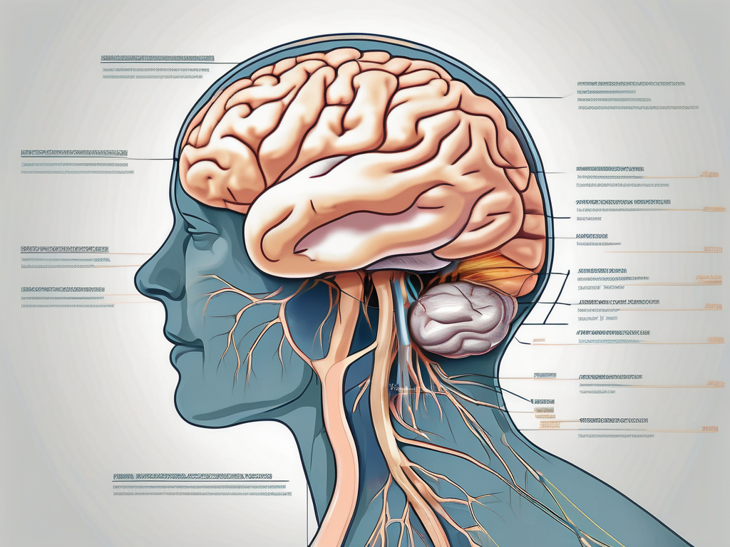
The abducens nerve originates in the pons, a region located in the brainstem. Specifically, it arises from the abducens nucleus, which is situated in the lower segment of the pons. This nucleus serves as the source of motor fibers that are primarily associated with the lateral rectus muscle of the eye.
The abducens nerve embarks on a long course, extending from the abducens nucleus to the orbit. As it travels through the skull, it passes through the cavernous sinus, a venous channel located on either side of the sella turcica, a bony structure at the base of the skull. Within the cavernous sinus, the abducens nerve is accompanied by other important structures, including the internal carotid artery and the oculomotor and trochlear nerves.
Upon reaching the orbit, the abducens nerve innervates the lateral rectus muscle. This muscle, as its name suggests, is responsible for the abduction of the eye. By contracting, the lateral rectus muscle pulls the eye outward, allowing it to gaze laterally. This movement is essential for binocular vision, as it enables both eyes to work together and provide optimal visual field coverage.
Function of the Abducens Nerve
The primary function of the abducens nerve is to control the lateral movement of the eye. This movement, facilitated by the lateral rectus muscle, allows the eye to gaze outward. By coordinating with other ocular motor nerves, such as the oculomotor and trochlear nerves, the abducens nerve ensures smooth and precise eye movements.
When the abducens nerve is functioning properly, it allows for the synchronized movement of both eyes. This coordination is crucial for various visual tasks, including tracking moving objects, maintaining visual fixation, and scanning the environment. Without the abducens nerve’s contribution, our ability to perceive the world around us would be significantly compromised.
However, like any other nerve, the abducens nerve is susceptible to damage or dysfunction. Conditions such as abducens nerve palsy, which is characterized by the inability to move the affected eye laterally, can result from trauma, infections, tumors, or other underlying medical conditions. Understanding the anatomy and function of the abducens nerve can aid in the diagnosis and management of such conditions.
In conclusion, the abducens nerve is a vital component of the ocular motor system, responsible for controlling the lateral movement of the eye. Its anatomy and function are intricately connected, allowing for precise eye movements and optimal visual field coverage. By delving into the complexities of the abducens nerve, we can appreciate the remarkable mechanisms that govern our ability to see and perceive the world around us.
The Synaptic Process of the Abducens Nerve
The synaptic process of the abducens nerve is a complex and fascinating phenomenon that involves the intricate interplay of various components. One crucial aspect of this process is the role of neurotransmitters, which play a vital role in facilitating communication between neurons.
The Role of Neurotransmitters
Neurotransmitters, such as acetylcholine, are key players in the synaptic process of the abducens nerve. Acetylcholine, the primary neurotransmitter involved in motor neuron signaling, is released from the abducens nerve terminal buttons. This release is carefully regulated and occurs in response to specific stimuli.
Upon reaching the neuromuscular junction, acetylcholine binds to receptors present on the muscle fibers. This binding event triggers a cascade of events that ultimately lead to muscle contraction and subsequent eye abduction. The binding of acetylcholine to its receptors initiates a series of biochemical reactions within the muscle fiber, resulting in the generation of force and movement.
It is worth noting that the release of neurotransmitters is a highly precise and tightly regulated process. The timing and quantity of neurotransmitter release are crucial for the proper functioning of the abducens nerve and the coordination of eye movements. Any disruption in this delicate balance can lead to motor impairments and difficulties in eye movement control.
Synaptic Transmission in the Abducens Nerve
The synaptic transmission process in the abducens nerve is a remarkable feat of biological engineering. It involves the transmission of nerve impulses across the synapse, the tiny gap between the abducens nerve and the muscle fibers it innervates.
When an action potential is generated in the abducens nucleus, it travels down the length of the nerve with astonishing speed. Within milliseconds, the nerve impulse reaches the neuromuscular junction, where it triggers the release of neurotransmitters. This rapid transmission ensures swift and accurate communication between the nerve and the muscle, allowing for precise control of eye movements.
The release of neurotransmitters at the neuromuscular junction is a highly coordinated process. It involves the fusion of synaptic vesicles containing neurotransmitters with the presynaptic membrane, followed by the diffusion of neurotransmitters into the synaptic cleft. Once in the synaptic cleft, neurotransmitters bind to receptors on the muscle fibers, initiating a series of events that ultimately lead to muscle contraction.
The synaptic transmission process in the abducens nerve is a remarkable example of the intricate mechanisms at play in the human body. It highlights the precision and efficiency with which our nervous system operates, allowing us to perform complex movements and tasks effortlessly.
The Pathway of the Abducens Nerve
The abducens nerve, also known as cranial nerve VI, plays a vital role in eye movement and coordination. Let’s explore the origin, course, and termination of this important nerve.
Origin and Course of the Abducens Nerve
The abducens nerve originates in the pons of the brainstem, specifically from the abducens nucleus. This nucleus serves as the starting point for the nerve fibers that will eventually control the lateral rectus muscle, responsible for moving the eye laterally.
From the abducens nucleus, the nerve fibers travel through the pons, a region located in the upper part of the brainstem. As the fibers make their way through the pons, they emerge from its ventral surface, forming the abducens nerve.
Once the abducens nerve is formed, it continues its journey through the skull. It passes through a small opening called the superior orbital fissure, which is located in the sphenoid bone. This opening provides a pathway for the nerve to reach its destination: the orbit.
Upon reaching the orbit, the abducens nerve innervates the lateral rectus muscle. This muscle is one of the six extraocular muscles responsible for controlling eye movement. By providing innervation to the lateral rectus muscle, the abducens nerve enables the eye to move laterally, allowing us to gaze to the side.
Termination of the Abducens Nerve
The abducens nerve terminates at the neuromuscular junction in the lateral rectus muscle. This junction is the point where the nerve fibers make contact with the muscle fibers, allowing for the transmission of signals and the initiation of muscle contraction.
The termination of the abducens nerve at the neuromuscular junction is crucial for the accurate control of the lateral rectus muscle. This precise control facilitates coordinated eye movement during lateral gaze, enabling us to shift our focus from one side to the other smoothly and effortlessly.
Proper alignment of both eyes is essential for binocular vision and depth perception. The interaction between the abducens nerve and the lateral rectus muscle ensures that both eyes move synchronously, allowing us to perceive the world in three dimensions.
In summary, the abducens nerve follows a fascinating pathway from its origin in the pons to its termination at the lateral rectus muscle. This pathway allows for the precise control and coordination of eye movement, contributing to our ability to explore and interact with our surroundings.
Disorders Related to the Abducens Nerve
The abducens nerve, also known as cranial nerve VI, plays a crucial role in eye movement. When this nerve is impaired or dysfunctional, it can lead to a condition known as abducens nerve palsy. This condition can arise due to various causes, and it is important to identify the underlying factor to determine the most appropriate treatment approach.
Causes of Abducens Nerve Palsy
Abducens nerve palsy can be caused by a range of factors. One common cause is trauma, such as a head injury or a direct blow to the eye area. Infections, such as meningitis or sinusitis, can also lead to the impairment of the abducens nerve. Additionally, tumors in the brain or surrounding structures can exert pressure on the nerve, leading to its dysfunction. Vascular disorders, including aneurysms or strokes, can also affect the abducens nerve. Furthermore, certain neurological conditions, such as multiple sclerosis, have been associated with abducens nerve palsy.
Understanding the cause of abducens nerve palsy is crucial in determining the appropriate treatment approach. Traumatic cases may require immediate medical intervention, while infections may be treated with antibiotics or antiviral medications. Tumors may necessitate surgical intervention or radiation therapy, depending on their nature and location. Vascular disorders may require specialized medical management to address the underlying issue, and neurological conditions may benefit from targeted therapies to manage symptoms and slow disease progression.
Symptoms and Diagnosis of Abducens Nerve Disorders
When the abducens nerve is affected, individuals may experience various symptoms. One common symptom is double vision, also known as diplopia. This occurs because the affected eye is unable to move properly, leading to misalignment with the unaffected eye. Strabismus, a condition characterized by the misalignment of the eyes, can also occur as a result of abducens nerve palsy. Individuals may notice that one eye deviates inward or outward, causing an imbalance in visual perception.
Diagnosing abducens nerve disorders involves a thorough examination by an ophthalmologist and a neurologist. The ophthalmologic examination assesses visual acuity, eye movements, and the alignment of the eyes. The neurologic evaluation aims to identify any underlying neurological conditions or abnormalities that may be contributing to the abducens nerve dysfunction. In some cases, imaging studies such as magnetic resonance imaging (MRI) or computed tomography (CT) scans may be necessary to assess the structural integrity of the nerve and surrounding structures.
By accurately diagnosing abducens nerve disorders, healthcare professionals can develop an appropriate treatment plan tailored to the individual’s needs. Treatment options may include corrective lenses, eye exercises, patching, or surgery, depending on the severity and underlying cause of the condition.
Treatment and Management of Abducens Nerve Disorders
The abducens nerve is responsible for controlling the movement of the eye, specifically the lateral rectus muscle. When this nerve becomes damaged or impaired, it can lead to a condition known as abducens nerve palsy. The treatment and management of abducens nerve disorders depend on various factors, including the underlying cause and the severity of the condition.
Therapeutic Approaches for Abducens Nerve Palsy
Conservative management is often the first line of treatment for abducens nerve palsy. This approach may involve the use of corrective lenses, prisms, or eye patches to help improve vision and alleviate symptoms. These non-invasive methods can be effective in mild cases of abducens nerve palsy, providing relief and allowing the individual to regain some level of normal eye movement.
However, in more severe or persistent cases, additional interventions may be necessary. Surgical options can include procedures such as recession or resection of the affected eye muscle, which can help restore proper alignment and improve eye movement. These surgical interventions are typically performed by ophthalmologists who specialize in eye muscle surgery.
In recent years, specialized therapies such as botulinum toxin injections have also been used to manage abducens nerve palsy. Botulinum toxin, commonly known as Botox, can be injected into the affected eye muscle to weaken its activity temporarily. This can help reduce the misalignment of the eyes and improve overall eye movement. However, it is important to note that the effects of botulinum toxin injections are temporary and may require repeated treatments.
It is crucial for individuals with abducens nerve palsy to consult with a qualified healthcare professional to determine the most appropriate treatment plan. A thorough evaluation of the underlying cause, the severity of the condition, and the individual’s medical history will help guide the treatment decisions. The healthcare professional may also consider other factors, such as the individual’s age, overall health, and lifestyle, when developing a personalized treatment plan.
Prognosis and Recovery from Abducens Nerve Disorders
The prognosis and recovery from abducens nerve disorders can vary significantly depending on several factors. The underlying cause of the condition, the extent of nerve damage, and the timeliness of intervention all play a role in determining the outcome.
In some cases, individuals with abducens nerve palsy may experience complete recovery with appropriate treatment and management. With time and proper rehabilitation, the affected eye muscle can regain its function, allowing for improved eye movement and alignment.
However, it is important to note that not all cases of abducens nerve disorders have a favorable prognosis. Some individuals may have long-lasting functional limitations, even with treatment. Factors such as the severity of nerve damage, the presence of other underlying medical conditions, and the individual’s overall health can influence the recovery process.
For individuals facing abducens nerve disorders, seeking professional medical advice is crucial. A healthcare professional with expertise in neurology or ophthalmology can provide a comprehensive evaluation and develop a tailored treatment plan. This may include a combination of therapies, such as physical therapy, occupational therapy, or vision therapy, to maximize the potential for recovery and improve the individual’s quality of life.
In conclusion, the abducens nerve is a critical component of the eye’s movement control system. Understanding its anatomy, function, synaptic process, pathway, and associated disorders can provide valuable insights into the intricate mechanisms at play. While this article offers a comprehensive overview of the treatment and management of abducens nerve disorders, it is important to consult with a healthcare professional for personalized advice and guidance tailored to individual circumstances.
Leave a Reply