The abducens nerve, also known as the sixth cranial nerve, plays a crucial role in eye movement. Understanding the anatomy and function of this nerve is essential in comprehending its significance and the potential disorders associated with it.
Understanding the Abducens Nerve
The abducens nerve is a motor nerve that originates from the pons, a part of the brainstem. It is responsible for controlling the lateral rectus muscle, which allows the eyeball to move outwards.
The abducens nerve, also known as the sixth cranial nerve, plays a crucial role in the complex system that governs eye movements. Without this nerve, our ability to move our eyes horizontally would be severely compromised.
Anatomy of the Abducens Nerve
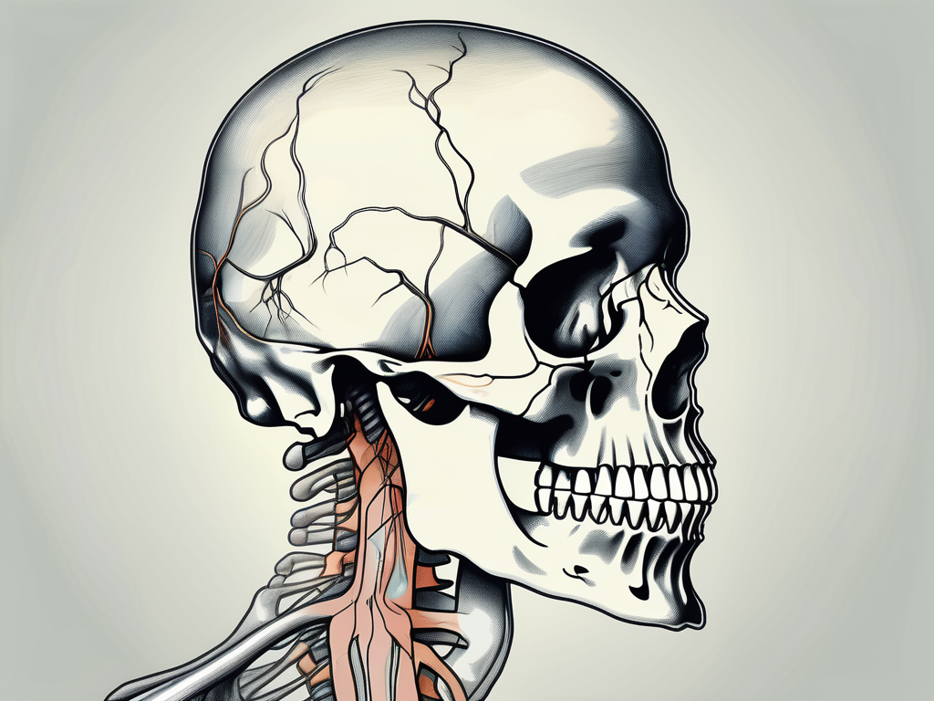
The abducens nerve emerges from the brainstem and passes through the cavernous sinus, a cavity located on the side of the skull. This path allows it to connect to the lateral rectus muscle.
As the abducens nerve traverses through the cavernous sinus, it is accompanied by other important structures such as the internal carotid artery and the oculomotor and trochlear nerves. This close proximity highlights the intricate nature of the cranial nerve network and the vital role it plays in coordinating eye movements.
The nerve’s fibers are primarily concentrated in the pons, which is located deep within the brainstem. From there, it extends downwards towards the midbrain and eventually exits the skull.
Upon exiting the skull, the abducens nerve travels along the lateral wall of the cavernous sinus before entering the orbit through the superior orbital fissure. It then reaches its destination, the lateral rectus muscle, where it exerts its control over eye movement.
Function of the Abducens Nerve
The primary function of the abducens nerve is to transmit signals from the brain to the lateral rectus muscle. This communication enables coordinated movement of both eyes for binocular vision.
Imagine trying to scan your surroundings without the ability to move your eyes horizontally. It would be incredibly challenging to explore the world around you and gather visual information efficiently. The abducens nerve ensures that this crucial eye movement is possible.
By controlling the lateral rectus muscle, the abducens nerve ensures that the eyes can move horizontally, contributing to activities such as scanning the environment, following objects, and maintaining proper alignment of both eyes.
Additionally, the abducens nerve works in conjunction with other cranial nerves, such as the oculomotor and trochlear nerves, to coordinate complex eye movements. This intricate interplay between multiple nerves allows us to perform tasks such as tracking moving objects, reading, and maintaining steady fixation on a target.
Furthermore, the abducens nerve is susceptible to various disorders and injuries that can disrupt its normal function. Conditions such as abducens nerve palsy, which is characterized by the inability to move the affected eye laterally, can result in double vision and difficulties with depth perception.
Understanding the abducens nerve and its role in eye movement highlights the incredible complexity and precision of the human body. The intricate network of cranial nerves working together ensures that our eyes can move harmoniously, allowing us to navigate the world with ease and clarity.
The Pathway of the Abducens Nerve
The abducens nerve follows a specific course within the skull, which ultimately determines its exit point.
The abducens nerve, also known as cranial nerve VI, plays a crucial role in eye movement. It innervates the lateral rectus muscle, which is responsible for abducting or moving the eye laterally away from the midline.
Origin of the Abducens Nerve
The abducens nerve originates from the abducens nucleus, located within the pons. This nucleus acts as a command center, controlling the movement of the lateral rectus muscle.
Within the abducens nucleus, motor neurons receive signals from various brain regions involved in eye movement coordination. These signals are integrated and processed, resulting in the generation of precise commands that will be transmitted to the lateral rectus muscle via the abducens nerve.
Once the necessary signals are generated within the abducens nucleus, the abducens nerve fibers begin their journey from the brainstem towards the skull.
Course of the Abducens Nerve
As the abducens nerve emerges from the brainstem, it travels through the cavernous sinus, a complex network of veins and nerves located on the side of the skull.
The cavernous sinus is a critical pathway for several cranial nerves and blood vessels. It is situated on either side of the sella turcica, a bony saddle-like structure that houses the pituitary gland.
Within the cavernous sinus, the abducens nerve runs alongside other important structures, including the internal carotid artery and the trochlear and oculomotor nerves. This close proximity allows for efficient communication and coordination between these structures, ensuring precise eye movements.
After traversing the cavernous sinus, the abducens nerve continues its descent through the base of the skull until it reaches its exit point.
The abducens nerve exits the skull through the superior orbital fissure, a narrow opening located in the sphenoid bone. This fissure serves as a passageway for several structures involved in eye movement and sensation, including the ophthalmic branch of the trigeminal nerve.
Once outside the skull, the abducens nerve branches out and innervates the lateral rectus muscle, enabling it to carry out its crucial role in moving the eye laterally.
Understanding the pathway of the abducens nerve is essential for diagnosing and treating various conditions that can affect eye movement. Damage or dysfunction of the abducens nerve can result in a condition known as abducens nerve palsy, which leads to impaired lateral eye movement and can cause double vision.
By studying the intricate course of the abducens nerve, researchers and healthcare professionals can gain valuable insights into the complex mechanisms underlying eye movement and develop effective treatments for related disorders.
The Abducens Nerve and the Skull
The Abducens Nerve’s Exit Point from the Skull
The exit point of the abducens nerve from the skull is known as the superior orbital fissure. This narrow opening is located at the base of the frontal bone, just above the orbit (eye socket).
As the abducens nerve traverses through the superior orbital fissure, it embarks on a remarkable journey that is vital for the intricate workings of the human eye. This exit point serves as a gateway, connecting the nerve’s origin within the skull to its destination within the orbit.
Once the abducens nerve passes through the superior orbital fissure, its fibers enter the orbit to innervate the lateral rectus muscle. This muscle, situated on the outer side of the eye, plays a pivotal role in the horizontal movement of the eyeball.
The abducens nerve’s meticulous pathway through the superior orbital fissure ensures that the lateral rectus muscle receives the necessary signals for coordinated eye movements. This intricate process allows us to effortlessly scan our surroundings, follow moving objects, and maintain visual stability.
Significance of the Exit Point
The exit point of the abducens nerve is not merely a geographical landmark; it holds immense significance in the realm of ocular function. This juncture marks the nerve’s transition from the protective confines of the skull to the dynamic environment of the orbit, where it fulfills its crucial role in eye movement.
Any disruption or compression of the abducens nerve at this exit point can have profound consequences, leading to various disorders that affect the functioning of the lateral rectus muscle and, consequently, eye movement. Conditions such as abducens nerve palsy, caused by trauma, inflammation, or compression, can result in the inability to abduct the affected eye, leading to double vision and impaired depth perception.
Understanding the intricate relationship between the abducens nerve and its exit point from the skull allows us to appreciate the delicate balance required for smooth eye movements. The superior orbital fissure serves as a gateway that connects the realms of anatomy and physiology, enabling the abducens nerve to fulfill its crucial role in our visual experience.
Disorders Related to the Abducens Nerve
The abducens nerve, also known as the sixth cranial nerve, plays a crucial role in eye movement. When this nerve is affected by disorders, it can lead to various symptoms and complications. Understanding the symptoms and treatment options for abducens nerve disorders is essential for proper management and care.
Symptoms of Abducens Nerve Disorders
Abducens nerve disorders can manifest in different ways, resulting in a range of symptoms. One of the most common symptoms is difficulties in moving the eye laterally, a movement known as abduction. This can lead to a limited range of eye movement and affect visual coordination.
Another common symptom of abducens nerve disorders is double vision, also known as diplopia. This occurs when the eyes are not properly aligned, causing the brain to receive conflicting images from each eye. Double vision can be disorienting and affect daily activities such as reading, driving, and even walking.
In addition to difficulties in eye movement and double vision, individuals with abducens nerve disorders may experience eye misalignment, where one eye may appear to be deviated or turned inward or outward. This misalignment can be noticeable and may cause self-consciousness or social discomfort.
Furthermore, the strain caused by improper eye coordination can lead to eye fatigue and headaches. The eyes work together to focus on objects, and when this coordination is disrupted, it can result in eye strain and discomfort. Headaches may also occur due to the extra effort required to compensate for the impaired eye movement.
Treatment Options for Abducens Nerve Disorders
If you suspect any issues with your abducens nerve or experience persistent symptoms, it is crucial to consult with a qualified healthcare professional or ophthalmologist. They can perform a thorough examination, evaluate your condition, and provide appropriate treatment options based on the findings.
Treatment for abducens nerve disorders depends on the underlying cause. In some cases, inflammation may be the culprit, and medications to reduce inflammation, such as corticosteroids, may be prescribed. These medications can help alleviate symptoms and promote healing of the affected nerve.
In other cases, abducens nerve disorders may be caused by compression or pressure on the nerve. Surgical intervention may be necessary to relieve this compression and restore proper nerve function. The specific surgical procedure will depend on the individual’s condition and the underlying cause of the disorder.
Additionally, if the abducens nerve disorder is secondary to an underlying condition, such as a brain tumor or aneurysm, the treatment plan will focus on addressing the primary condition. This may involve a combination of surgery, radiation therapy, or other targeted therapies to manage the underlying cause and alleviate the associated abducens nerve disorder.
Furthermore, certain therapies may be recommended to address specific underlying conditions that contribute to abducens nerve disorders. These therapies can include vision exercises, eye patches, prisms, or other interventions aimed at improving eye coordination and alignment.
In conclusion, abducens nerve disorders can have a significant impact on eye movement and coordination. Recognizing the symptoms and seeking appropriate medical attention is crucial for accurate diagnosis and effective treatment. With the right interventions, individuals with abducens nerve disorders can experience improved eye function and quality of life.
Frequently Asked Questions about the Abducens Nerve
Common Misconceptions about the Abducens Nerve
One common misconception is that abducens nerve disorders are solely caused by eye strain or fatigue. While prolonged eye strain can contribute to eye fatigue, abducens nerve disorders often have complex underlying causes.
It is essential to seek professional medical advice to accurately diagnose and treat any potential issues with the abducens nerve.
Abducens nerve disorders can be caused by various factors, including trauma to the head or eye, infections, tumors, or underlying medical conditions such as diabetes or multiple sclerosis. These conditions can affect the function of the abducens nerve and lead to symptoms such as double vision, difficulty moving the eyes laterally, or even complete paralysis of eye movement.
Diagnosing abducens nerve disorders requires a comprehensive evaluation by a healthcare professional. They may perform a physical examination, review medical history, and order additional tests such as imaging studies or nerve conduction tests to determine the underlying cause and develop an appropriate treatment plan.
Recent Research Findings on the Abducens Nerve
Ongoing research continues to shed light on the intricate details of the abducens nerve’s anatomy, physiology, and associated disorders. New studies explore potential therapeutic approaches, improved diagnostic techniques, and a deeper understanding of this critical nerve’s role in eye movement.
Researchers are investigating the molecular mechanisms that regulate the development and function of the abducens nerve. They aim to identify specific genes and proteins involved in its formation and maintenance, which could potentially lead to targeted therapies for abducens nerve disorders.
Furthermore, recent studies have focused on the relationship between the abducens nerve and other cranial nerves. Understanding the interconnectedness of these nerves can provide insights into the complex coordination required for precise eye movements.
Advancements in imaging technology, such as high-resolution magnetic resonance imaging (MRI) and optical coherence tomography (OCT), have allowed researchers to visualize the abducens nerve in more detail. These imaging techniques help identify structural abnormalities or damage to the nerve, aiding in the diagnosis and monitoring of abducens nerve disorders.
Stay informed about the latest research findings through reputable medical journals and consult with medical professionals to ensure up-to-date and accurate information.
It is important to note that while research is advancing our understanding of the abducens nerve, there is still much to learn. Continued research efforts will contribute to improved treatments and outcomes for individuals with abducens nerve disorders.
In conclusion, the abducens nerve exits the skull through the superior orbital fissure, marking its transition from the skull to the orbit. Understanding the anatomy, function, and potential disorders related to this nerve is essential for comprehending its significant role in eye movement. If you have concerns or experience symptoms related to your abducens nerve, it is crucial to consult with a healthcare professional for proper evaluation and guidance.
Leave a Reply