The abducens nerve, also known as the sixth cranial nerve, plays a crucial role in eye movement. Located within the brainstem, this nerve is responsible for controlling the lateral rectus muscle of the eye. Understanding the anatomy, function, and disorders related to the abducens nerve can provide valuable insights into its importance in daily life.
Understanding the Abducens Nerve
The abducens nerve is a fascinating component of the human nervous system. Its role in eye movement and alignment is crucial for our ability to perceive the world around us. Let’s delve deeper into the definition, function, and anatomy of this remarkable cranial nerve.
Definition and Function of the Abducens Nerve
The abducens nerve, also known as cranial nerve VI, is one of the twelve cranial nerves originating from the brainstem. It emerges from the pons, a region in the brainstem responsible for relaying signals between the cerebral cortex and the spinal cord.
Extending from the pons towards the eye, the abducens nerve plays a vital role in eye movement. It primarily innervates the lateral rectus muscle, which is responsible for moving the eye away from the midline of the body. This movement, known as abduction, allows us to shift our gaze laterally and maintain proper alignment of the eyes.
Anatomy of the Abducens Nerve
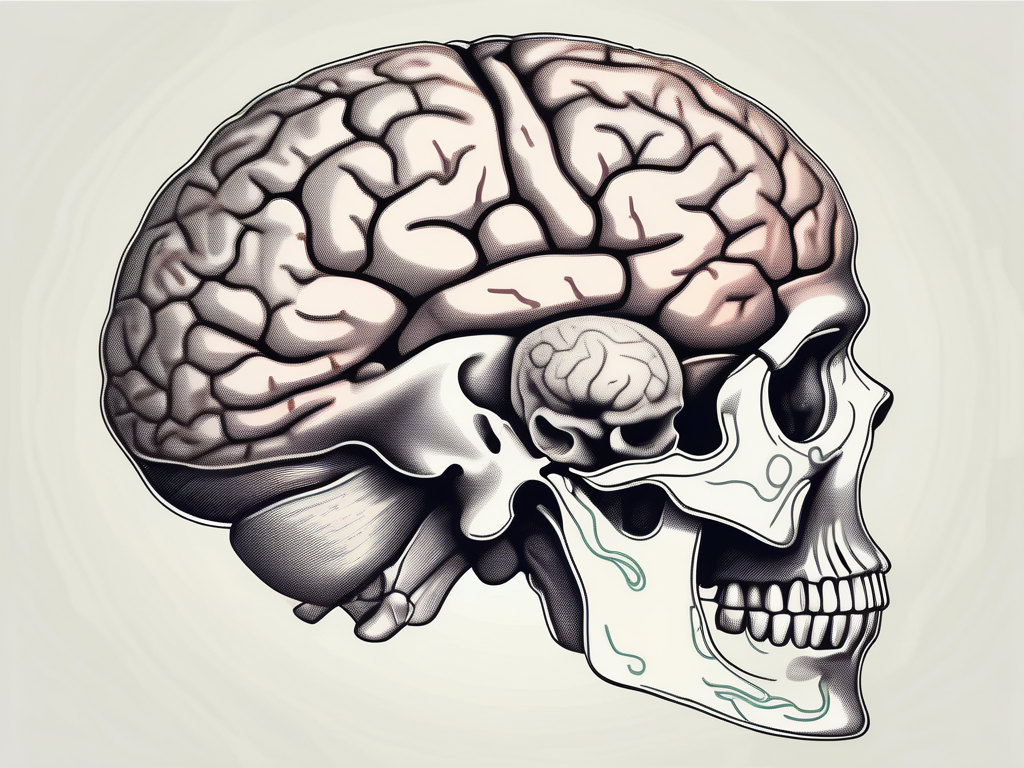
The anatomical pathway of the abducens nerve is a marvel of precision and complexity. After exiting the brainstem, it embarks on a journey through various structures in the head.
Passing through the cavernous sinus, a space located behind the eyes, the abducens nerve navigates its way towards the orbit. The cavernous sinus is a critical region that houses several important structures, including blood vessels and other cranial nerves.
Once inside the orbit, the abducens nerve reaches its destination – the lateral rectus muscle. This muscle, situated within the bony cavity surrounding the eyeball, receives innervation from the abducens nerve. The precise control exerted by this nerve allows for smooth and coordinated eye movements, ensuring our visual perception is accurate and reliable.
Understanding the intricate anatomy and function of the abducens nerve provides us with a deeper appreciation for the complexity of the human body. The coordination required for eye movements is a testament to the remarkable capabilities of our nervous system.
The Pathway of the Abducens Nerve
The abducens nerve, also known as the sixth cranial nerve, plays a crucial role in eye movement. It originates from the abducens nucleus, a small group of nerve cells located within the pons, a part of the brainstem. This nucleus serves as the starting point for the abducens nerve’s journey.
Once the abducens nerve emerges from the abducens nucleus, it begins its course along a specific pathway. This pathway takes it on a fascinating journey, crossing over the midline of the brainstem. This crossing, known as the decussation, ensures that the abducens nerve connects with the appropriate structures on the opposite side of the brainstem.
As the abducens nerve continues its journey, it eventually reaches its termination point in the lateral rectus muscle of the eye. The lateral rectus muscle is one of the six extraocular muscles responsible for controlling eye movement. Specifically, the abducens nerve innervates the lateral rectus muscle, allowing it to contract and move the eye laterally, away from the midline of the face.
Relation to Other Cranial Nerves
The abducens nerve does not work alone in the complex task of eye movement. It shares proximity and connections with other cranial nerves that are also involved in this intricate process. One of the most significant connections is with the oculomotor nerve, the third cranial nerve.
The oculomotor nerve is responsible for controlling most of the other ocular muscles, including the superior rectus, inferior rectus, and medial rectus muscles. These muscles work together to move the eye in various directions, allowing for smooth and accurate tracking of objects in the visual field.
The abducens nerve and the oculomotor nerve work in harmony to coordinate eye movements. While the abducens nerve controls the lateral rectus muscle, which moves the eye laterally, the oculomotor nerve controls the other ocular muscles, enabling vertical and medial movements. This coordinated effort ensures that the eyes can accurately follow objects and maintain visual perception.
Understanding the pathway and relationships of the abducens nerve provides valuable insights into the complex mechanisms that govern eye movement. The intricate interplay between cranial nerves highlights the remarkable precision and coordination required for our eyes to function optimally.
Role of the Abducens Nerve in Eye Movement
The abducens nerve, also known as the sixth cranial nerve, plays a crucial role in controlling eye movement. It is responsible for transmitting signals to the lateral rectus muscle, which enables the eye to move away from the midline, a process known as abduction. This movement is essential for various activities that require peripheral vision, such as scanning the environment, reading, and driving.
When the abducens nerve functions properly, it allows for precise coordination between the eyes, ensuring smooth and accurate eye movements. However, when there is a dysfunction or impairment in the abducens nerve, individuals may experience a lack of coordination in their eye movements, leading to difficulties in visual perception.
Controlling Lateral Rectus Muscle
The abducens nerve’s primary responsibility lies in controlling the lateral rectus muscle. This muscle is one of the six extraocular muscles responsible for moving the eye. By transmitting signals to the lateral rectus muscle, the abducens nerve enables the eye to move laterally, away from the midline of the face.
Imagine a scenario where you are reading a book. As you progress through the text, your eyes need to move from left to right, scanning each line. This lateral movement is made possible by the abducens nerve, which ensures that the lateral rectus muscle contracts and pulls the eye outward, allowing you to smoothly track the words on the page.
Similarly, when driving, you rely on your peripheral vision to be aware of your surroundings. The abducens nerve plays a vital role in enabling your eyes to quickly shift their focus from the road ahead to the side mirrors, ensuring that you have a comprehensive view of the traffic around you.
Involvement in Gaze Mechanism
The abducens nerve is not only responsible for controlling the lateral rectus muscle but also plays a significant role in the complex gaze mechanism. The gaze mechanism is a network of neural pathways that allows us to fixate on objects of interest and track their movements.
Working in conjunction with other cranial nerves, the abducens nerve ensures precise coordination between the eyes, allowing for various types of eye movements. These include smooth pursuit, which involves smoothly tracking a moving object, saccades, which are rapid eye movements between fixation points, and vestibular ocular reflexes, which help stabilize our gaze during head movements.
Imagine watching a bird flying across the sky. As it moves, your eyes need to smoothly track its flight path. This is made possible by the coordinated efforts of the abducens nerve and other cranial nerves, ensuring that your eyes move in perfect synchronization to keep the bird in focus.
In addition to smooth pursuit, the abducens nerve also plays a crucial role in saccades. Saccades allow our eyes to rapidly shift their focus from one point to another, enabling us to explore our environment and quickly gather visual information. Dysfunction of the abducens nerve can disrupt the precise coordination required for saccades, leading to difficulties in shifting our gaze efficiently.
Furthermore, the abducens nerve contributes to vestibular ocular reflexes, which help stabilize our gaze during head movements. When we turn our heads, the abducens nerve ensures that our eyes move in the opposite direction, allowing us to maintain a stable visual field. Dysfunction of the abducens nerve can result in gaze instability, making it challenging to maintain a clear and focused view when our head is in motion.
In conclusion, the abducens nerve plays a crucial role in controlling eye movement. By transmitting signals to the lateral rectus muscle and participating in the complex gaze mechanism, it enables us to have precise and coordinated eye movements. Dysfunction of the abducens nerve can lead to difficulties in visual perception, coordination, and gaze stability. Understanding the role of the abducens nerve helps us appreciate the intricate mechanisms that allow us to explore and interact with the visual world around us.
Disorders Related to the Abducens Nerve
The abducens nerve, also known as the sixth cranial nerve, plays a crucial role in the control of eye movements. When this nerve is affected by various disorders, it can lead to a condition called abducens nerve palsy. This condition is characterized by the paralysis or weakness of the muscles that control the lateral movement of the eye.
Causes of Abducens Nerve Palsy
Abducens nerve palsy can result from a variety of causes. One common etiology is the compression of the nerve due to lesions or tumors in the brainstem or cavernous sinus. These growths can exert pressure on the abducens nerve, disrupting its normal function and leading to palsy.
In addition to structural abnormalities, certain medical conditions can also contribute to abducens nerve dysfunction. For example, individuals with diabetes may be at an increased risk of developing nerve damage, including damage to the abducens nerve. Similarly, hypertension, or high blood pressure, can affect the blood vessels supplying the nerve, compromising its function. Trauma, such as head injuries or fractures, can also lead to abducens nerve palsy.
Symptoms and Diagnosis of Abducens Nerve Disorders
Individuals with abducens nerve disorders may experience a range of symptoms. One common symptom is double vision, also known as diplopia. This occurs when the affected eye is unable to align properly with the other eye, resulting in overlapping images. Another symptom is uncoordinated eye movements, where the affected eye may not move in sync with the unaffected eye, leading to impaired vision and difficulty focusing.
Diagnosing abducens nerve disorders typically involves a comprehensive evaluation by a healthcare professional. This evaluation includes taking a thorough medical history to identify any underlying conditions or previous injuries that may contribute to the nerve dysfunction. A physical examination is also conducted, which may involve assessing the movement of the eyes and looking for any signs of weakness or paralysis. In some cases, diagnostic imaging tests, such as magnetic resonance imaging (MRI) or computed tomography (CT) scans, may be necessary to visualize the structures surrounding the abducens nerve and identify any potential causes of palsy.
Prompt and accurate diagnosis of abducens nerve disorders is vital for appropriate management. Depending on the underlying cause, treatment options may vary. In some cases, addressing the underlying medical condition, such as controlling blood sugar levels in diabetes or managing hypertension, can help improve abducens nerve function. Physical therapy and eye exercises may also be recommended to strengthen the muscles around the affected eye and improve coordination. In more severe cases, surgical intervention may be necessary to relieve compression on the nerve or address any structural abnormalities.
Treatment and Management of Abducens Nerve Disorders
Abducens nerve disorders can significantly impact a person’s quality of life and daily functioning. The treatment and management of these disorders vary depending on the underlying cause and severity of the condition. In this article, we will explore some of the medical interventions, rehabilitation, and therapy options available for individuals with abducens nerve disorders.
Medical Interventions
When it comes to treating abducens nerve disorders, the first step is to identify the underlying cause. This is crucial in determining the most appropriate course of action. In cases where the disorder is caused by nerve compression or the presence of tumors, surgical intervention may be necessary to relieve the pressure on the nerve.
Surgery can help alleviate the symptoms associated with abducens nerve disorders and restore normal functioning. During the procedure, the surgeon carefully removes any obstructions or tumors that may be compressing the nerve, allowing it to function properly once again. It is important to note that surgical intervention is typically considered a last resort and is only recommended when other treatment options have been exhausted or when the condition poses a significant risk to the individual’s health.
In addition to surgical interventions, medications can also be prescribed to manage associated inflammation or swelling. Anti-inflammatory drugs or corticosteroids may be used to reduce inflammation and provide relief from symptoms. However, it is crucial to consult with a healthcare professional before starting any medication to ensure it is safe and appropriate for your specific condition.
Rehabilitation and Therapy Options
While medical interventions are essential for treating abducens nerve disorders, rehabilitation and therapy options also play a vital role in the management and recovery process. These therapies focus on improving eye coordination, functionality, and overall quality of life.
Visual therapy and rehabilitation techniques are often recommended for individuals with abducens nerve palsy. These therapies are conducted under the guidance of trained professionals who specialize in eye-related conditions. The primary goal of visual therapy is to strengthen the ocular muscles, enhance visual tracking, and improve overall eye movement control.
During visual therapy sessions, individuals may engage in various exercises and activities designed to target specific areas of concern. These exercises can range from simple eye movements to more complex tasks that challenge the individual’s visual abilities. Over time, consistent practice and guidance from the therapist can lead to significant improvements in eye coordination and functionality.
In addition to visual therapy, physical therapy and occupational therapy may also form part of the rehabilitation process for individuals with abducens nerve disorders. Physical therapy focuses on improving overall strength, flexibility, and balance, which can indirectly benefit eye coordination and functionality. Occupational therapy, on the other hand, addresses any additional functional concerns that may arise from the condition, such as difficulties with daily activities or work-related tasks.
It is important to note that every individual’s treatment plan will be tailored to their specific needs and goals. Therefore, it is crucial to consult with a healthcare professional or specialist who can provide personalized advice and guidance based on your unique circumstances.
The Abducens Nerve: A Comprehensive Overview
The abducens nerve, also known as the sixth cranial nerve, plays a crucial role in the intricate system of eye movement control. It is responsible for innervating the lateral rectus muscle, which facilitates lateral gaze. Understanding the anatomy, function, and disorders related to the abducens nerve is essential in recognizing its significance in daily life.
Anatomy of the Abducens Nerve
The abducens nerve originates from the pons, a region of the brainstem. It emerges from the brainstem and travels through the cavernous sinus, a cavity located behind the eye socket. From there, it enters the orbit and innervates the lateral rectus muscle, which is responsible for moving the eye outward.
The abducens nerve consists of motor fibers that control the lateral rectus muscle and allow for horizontal eye movements. These movements are essential for tasks such as reading, driving, and maintaining visual awareness of our surroundings.
Function of the Abducens Nerve
The primary function of the abducens nerve is to control the lateral rectus muscle, which is responsible for abduction of the eye. Abduction refers to the movement of the eye away from the midline of the face, allowing us to look to the side.
When the abducens nerve is functioning properly, it coordinates with other cranial nerves and muscles to ensure smooth and coordinated eye movements. This enables us to shift our gaze from one point to another, track moving objects, and explore our visual environment.
Disorders Related to the Abducens Nerve
Various disorders can affect the abducens nerve, leading to abnormalities in eye movement control. One common condition is abducens nerve palsy, which occurs when the nerve is damaged or compressed. This can result in a weakened or paralyzed lateral rectus muscle, leading to a limited ability to move the eye laterally.
Other conditions that can affect the abducens nerve include tumors, infections, and certain neurological disorders. These conditions may cause symptoms such as double vision, eye misalignment, and difficulty with lateral gaze.
Seeking Professional Help
If you experience any symptoms or concerns related to your eye movements, it is crucial to consult with a qualified healthcare professional. They can provide an accurate diagnosis, expert guidance, and tailored management strategies to address any issues related to the abducens nerve.
Early detection and intervention are key in managing disorders of the abducens nerve. Prompt medical attention can help prevent further complications and improve overall eye movement function.
Remember, the abducens nerve plays a vital role in our daily lives, allowing us to perform essential tasks and navigate our visual world. Taking care of our eye health and seeking appropriate medical care when needed ensures that we can continue to enjoy clear and effortless eye movements.
Leave a Reply