The anatomy of the eye is a complex and fascinating subject. Understanding how the various structures work together to enable vision is crucial in appreciating the mechanisms behind eye movement. One important component of this intricate system is the group of six extrinsic eye muscles. These muscles play a crucial role in directing the movement of our eyes, allowing us to focus on objects and explore our surroundings.
Understanding the Anatomy of the Eye
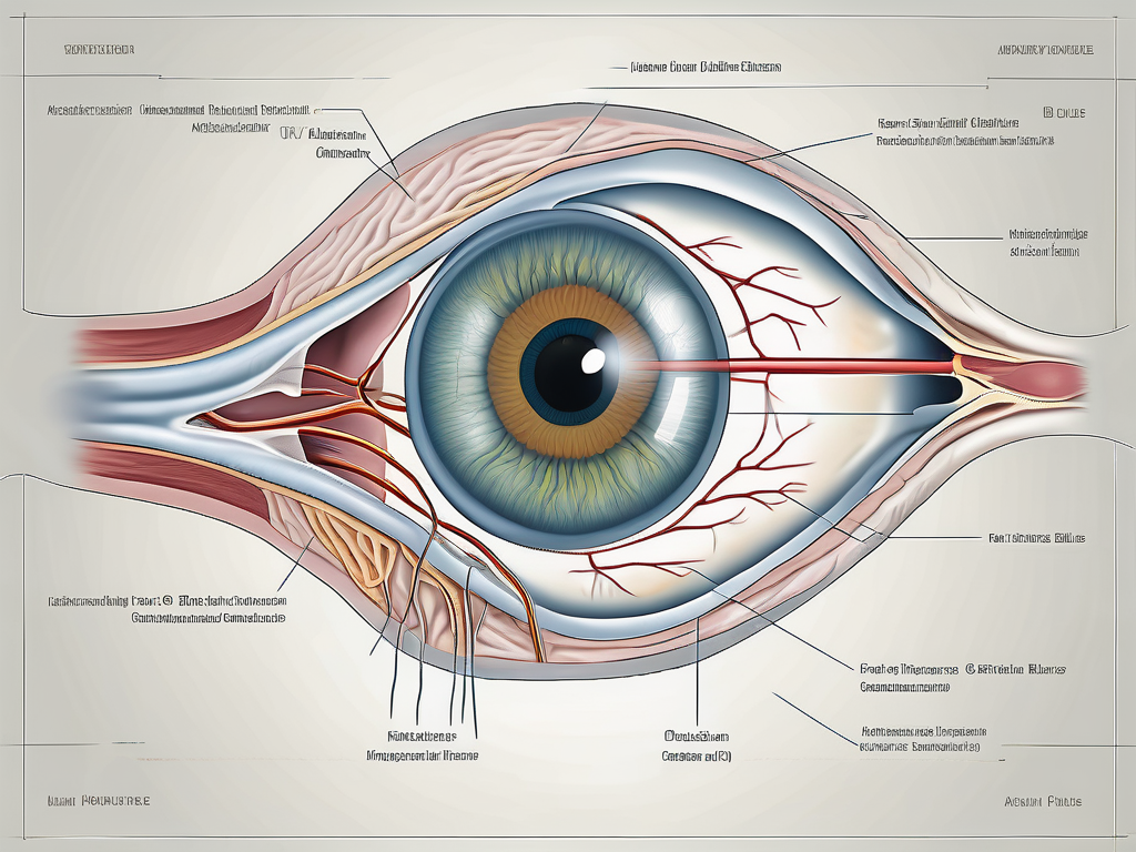
Before delving into the specifics of the abducens nerve and the inferior oblique muscle, it is essential to have a basic understanding of the anatomy of the eye. The eye is a delicate organ, protected by the orbit, also known as the eye socket. Within the orbit, the eye itself is surrounded by various tissues, including muscles, nerves, blood vessels, and connective tissue.
The globe of the eye is made up of several layers, including the cornea, iris, lens, and retina. These structures work together to capture visual stimuli and transmit them to the brain, where the information is processed and interpreted. The extrinsic eye muscles, including the inferior oblique muscle, play a crucial role in facilitating this process.
Additionally, the eye is supported by a network of blood vessels that provide essential nutrients and oxygen to its various components. The ophthalmic artery, a branch of the internal carotid artery, supplies blood to the eye, ensuring its proper function and health.
The Role and Function of Extrinsic Eye Muscles
The extrinsic eye muscles are responsible for the voluntary movement of the eye. These muscles are attached to the outer surface of the eye and control its movements. By contracting and relaxing, the extrinsic eye muscles allow us to shift our gaze and track objects in our field of vision.
The six extrinsic eye muscles are the superior rectus, inferior rectus, medial rectus, lateral rectus, superior oblique, and inferior oblique. Each muscle has a specific role in eye movement, working together to create coordinated motions. In healthy individuals, these muscles work seamlessly and harmoniously to provide smooth, accurate eye movements.
Moreover, the extrinsic eye muscles are surrounded by a layer of connective tissue called the Tenon’s capsule. This capsule provides support and protection to the muscles, ensuring their proper functioning and preventing any damage or displacement.
The Importance of Nerve Innervation in Eye Movement
Eye movement is a complex process that relies heavily on the precise coordination of the extrinsic eye muscles. This coordination is achieved through the intricate interplay between these muscles and the nerves that innervate them. Each extrinsic eye muscle is controlled by a specific cranial nerve, ensuring the proper functioning of eye movements.
One of these cranial nerves is the abducens nerve, also known as cranial nerve VI (CN VI). The abducens nerve innervates the lateral rectus muscle, which is responsible for abducting or outwardly rotating the eye. This important nerve supplies the lateral rectus muscle with the necessary signals to contract and move the eye laterally.
In addition to the abducens nerve, the oculomotor nerve (cranial nerve III) and the trochlear nerve (cranial nerve IV) also play significant roles in eye movement. The oculomotor nerve innervates four of the six extrinsic eye muscles, namely the superior rectus, inferior rectus, medial rectus, and inferior oblique muscles. The trochlear nerve, on the other hand, innervates the superior oblique muscle, which is responsible for downward and inward eye movements.
The precise coordination of these cranial nerves and their corresponding muscles allows for the smooth and accurate movement of the eye in various directions, enabling us to explore our visual environment with ease.
The Abducens Nerve (CN VI) Explained
The abducens nerve, as mentioned earlier, is the sixth cranial nerve and plays a significant role in eye movement. It arises from the brainstem and travels a complex pathway to reach its target muscle, the lateral rectus. Understanding the origin and pathway of this nerve is crucial in comprehending its importance in eye movement.
The abducens nerve originates from the lower portion of the pons, a region in the brainstem. This region is responsible for relaying signals between the cerebral cortex and the spinal cord. The pons also plays a role in regulating vital functions such as breathing and sleeping. From its origin, the abducens nerve travels anteriorly through the brainstem, passing through various structures that are involved in coordinating motor functions.
As the abducens nerve continues its journey, it exits the skull through a small bony canal called the superior orbital fissure. This canal serves as a protective pathway for the nerve, shielding it from potential damage. Once outside the skull, the abducens nerve enters the orbit, the bony cavity that houses the eyeball, and courses towards its target muscle, the lateral rectus.
Within the orbit, the abducens nerve runs alongside other important structures, such as blood vessels and connective tissue. These structures provide essential support and nourishment to the nerve, ensuring its proper functioning. The abducens nerve’s pathway within the orbit is carefully orchestrated to ensure precise control over eye movement.
As it reaches the lateral rectus muscle, the abducens nerve branches into numerous smaller nerve fibers, each of which communicates with individual muscle fibers. This intricate branching allows for fine motor control, enabling the eye to move laterally with precision. The coordinated contraction and relaxation of the lateral rectus muscle, under the influence of the abducens nerve, allow for smooth and accurate eye movements.
Clinical Significance of the Abducens Nerve
The abducens nerve is vulnerable to various pathological conditions that can affect its normal functioning. Conditions such as nerve compression, trauma, inflammation, or tumors can disrupt the nerve’s ability to transmit signals to the lateral rectus muscle effectively.
When the abducens nerve is affected, it can lead to a condition known as abducens nerve palsy. Abducens nerve palsy results in the paralysis or weakness of the lateral rectus muscle, impairing the eye’s ability to abduct. This can cause double vision, difficulty in focusing on distant objects, and eye misalignment, which can significantly impact a person’s quality of life.
Diagnosing and treating abducens nerve palsy requires a thorough understanding of the nerve’s anatomy and function. Medical professionals use various diagnostic techniques, such as imaging studies and neurological examinations, to assess the extent of nerve damage and determine the underlying cause. Treatment options may include medication, physical therapy, or surgical interventions, depending on the specific circumstances.
Research continues to shed light on the intricate workings of the abducens nerve and its role in eye movement. Scientists and medical professionals strive to develop innovative therapies and interventions to improve outcomes for individuals with abducens nerve-related conditions. By unraveling the complexities of this vital cranial nerve, they aim to enhance our understanding of the human visual system and pave the way for advancements in ophthalmology and neurology.
The Inferior Oblique Muscle and its Innervation
While the abducens nerve primarily innervates the lateral rectus muscle, it is worth exploring the relationship between this nerve and other extrinsic eye muscles. One such muscle is the inferior oblique muscle, which plays an important role in eye movement and stability.
Anatomy and Function of the Inferior Oblique Muscle
The inferior oblique muscle is one of the six extrinsic eye muscles and shares a close anatomical relationship with the abducens nerve. Located on the inferior surface of the eye, the inferior oblique muscle is responsible for elevating the eye and helping with horizontal gaze.
Its origin can be traced back to the maxillary bone, near the anterior and inferior orbital rim. From there, it extends posteriorly and laterally, attaching to the posterior part of the eyeball. This unique positioning allows the inferior oblique muscle to exert its influence on eye movement.
When the inferior oblique muscle contracts, it causes the eye to rotate upward and outward. This movement is particularly important during certain eye movements, such as when looking up and away from the midline. It also helps to counteract the actions of other eye muscles, ensuring precise control over eye position.
Unlike the lateral rectus muscle, which is primarily innervated by the abducens nerve, the inferior oblique muscle is innervated by another cranial nerve known as the oculomotor nerve (CN III). The oculomotor nerve is responsible for controlling several extrinsic eye muscles, including the superior rectus, inferior rectus, and medial rectus, in addition to the inferior oblique.
The oculomotor nerve originates from the midbrain and travels through the cavernous sinus before entering the orbit. Once inside the orbit, it divides into several branches, one of which supplies the inferior oblique muscle with the necessary motor innervation.
The Relationship between the Inferior Oblique and the Abducens Nerve
While the abducens nerve does not directly innervate the inferior oblique muscle, the two structures work in concert to achieve coordinated eye movements. The abducens nerve plays a crucial role in mediating horizontal eye movements, which are fundamental for visual exploration and stability.
When we look towards the side, the abducens nerve sends signals to the lateral rectus muscle, causing it to contract and pull the eye laterally. At the same time, the inferior oblique muscle is activated to counteract the rotational forces generated by the lateral rectus muscle. This coordinated effort ensures that the eye moves smoothly and accurately, allowing us to track objects of interest.
Furthermore, the abducens nerve and the inferior oblique muscle work together to maintain eye stability. The inferior oblique muscle helps to prevent excessive rotation of the eye, ensuring that it remains aligned with the visual field. This stability is crucial for maintaining clear and focused vision.
In summary, while the abducens nerve primarily innervates the lateral rectus muscle, it is important to recognize the role of the inferior oblique muscle in eye movement and stability. The close anatomical relationship between these structures allows for coordinated eye movements and precise control over eye position. Understanding the innervation and function of the inferior oblique muscle enhances our knowledge of the complex mechanisms involved in visual exploration and gaze control.
Disorders Related to the Abducens Nerve and Inferior Oblique Muscle
When discussing the abducens nerve and the inferior oblique muscle, it is important to understand the potential disorders that can affect these structures. Seeking medical attention and proper diagnosis is essential for addressing these conditions effectively.
The abducens nerve, also known as the sixth cranial nerve, plays a crucial role in eye movement. It innervates the lateral rectus muscle, which is responsible for moving the eye outward. Any disruption or damage to this nerve can result in a condition known as abducens nerve palsy.
Symptoms and Diagnosis of Abducens Nerve Palsy
Abducens nerve palsy can lead to various symptoms, all of which can significantly impact an individual’s daily activities and overall quality of life. One of the most common symptoms is double vision, also known as diplopia. This occurs because the affected eye is unable to move outward properly, causing the images to overlap.
In addition to double vision, individuals with abducens nerve palsy may also experience eye misalignment, known as strabismus. This misalignment can cause the eyes to appear crossed or deviated, leading to aesthetic concerns and potential social discomfort.
Another symptom of abducens nerve palsy is difficulty in focusing on distant objects. This can make tasks such as driving or reading signs challenging and can significantly affect an individual’s independence and ability to perform daily activities.
If you experience any of these symptoms or notice any changes in your eye movements, it is crucial to consult with an eye care professional. They can perform a comprehensive examination to assess the function of the abducens nerve and other relevant structures, providing an accurate diagnosis and appropriate treatment options.
Treatment Options for Disorders of the Inferior Oblique Muscle
While the abducens nerve primarily innervates the lateral rectus muscle, disorders affecting the inferior oblique muscle can also impact eye movement. The inferior oblique muscle is responsible for elevating and rotating the eye. Conditions such as muscle weakness, abnormalities, or misalignment can cause visual disturbances and discomfort.
If you suspect any issues with your inferior oblique muscle, it is advisable to consult with an experienced ophthalmologist. They can perform a thorough evaluation, which may include imaging studies and other diagnostic tests, to determine the underlying cause of the problem.
Treatment options for disorders of the inferior oblique muscle depend on the specific condition and its severity. In some cases, exercises and eye muscle training may be recommended to strengthen the muscle and improve its function. Corrective lenses, such as prism glasses, can also help alleviate visual disturbances caused by muscle abnormalities.
In more severe cases, surgical intervention may be necessary. Surgery can involve adjusting the position or tension of the inferior oblique muscle to correct misalignment or weakness. The specific surgical approach will depend on the individual’s condition and the ophthalmologist’s expertise.
Overall, disorders related to the abducens nerve and inferior oblique muscle can have a significant impact on an individual’s visual function and quality of life. Seeking timely medical attention and appropriate treatment can help manage these conditions effectively and improve overall eye health and well-being.
The Impact of Abducens Nerve on Eye Movement
In conclusion, the abducens nerve plays a crucial role in horizontal eye movement, facilitating the outward rotation of the eye. Through its close relationship with the lateral rectus muscle, the abducens nerve ensures the coordinated and synchronized motion of the eyes, allowing for accurate visual exploration.
The Role of the Abducens Nerve in Horizontal Eye Movement
Horizontal eye movement is essential for scanning the environment, tracking moving objects, and maintaining stability during locomotion. The abducens nerve provides the essential neural signals that enable precise control and coordination of this critical eye movement.
When we look around, our eyes move in a smooth and coordinated manner, thanks to the abducens nerve. This nerve originates from the brainstem and innervates the lateral rectus muscle, which is responsible for the outward rotation of the eye. Without the abducens nerve, our eyes would not be able to move horizontally, limiting our ability to explore our surroundings and gather visual information.
Interestingly, the abducens nerve is the sixth cranial nerve and is one of the longest nerves in the body. It travels a complex pathway from the brainstem, passing through various structures before reaching the lateral rectus muscle. This intricate pathway ensures that the abducens nerve delivers the necessary signals for precise eye movements.
How the Abducens Nerve Contributes to Vision Stability
By working in concert with other extrinsic eye muscles, such as the inferior oblique, the abducens nerve contributes to the stability of our vision. The coordinated movements of the eyes allow for clear, focused vision, providing us with a comprehensive understanding of our surroundings.
Imagine trying to read a book or follow a moving object without the ability to move your eyes horizontally. It would be challenging to maintain a stable image and track the object accurately. The abducens nerve ensures that our eyes can move smoothly and effortlessly, allowing us to perceive the world around us with clarity and precision.
Furthermore, the abducens nerve also plays a role in maintaining eye alignment. When the abducens nerve is functioning correctly, both eyes move together, ensuring that they are always aligned and working in harmony. This alignment is crucial for binocular vision, depth perception, and overall visual comfort.
While understanding the intricate details of nerve innervation and muscle function is crucial in appreciating the complexities of eye movement, it is essential to remember that individual circumstances may vary. If you have any concerns about your eye health or experience any symptoms, it is always recommended to consult with a qualified healthcare professional for an accurate diagnosis and appropriate management.
Leave a Reply